

Changes of statistical structural fluctuations unveils an early compacted degraded stage of PNS myelin
- PMID: 24962806
- PMCID: PMC4069690
- DOI: 10.1038/srep05430
Changes of statistical structural fluctuations unveils an early compacted degraded stage of PNS myelin
Abstract
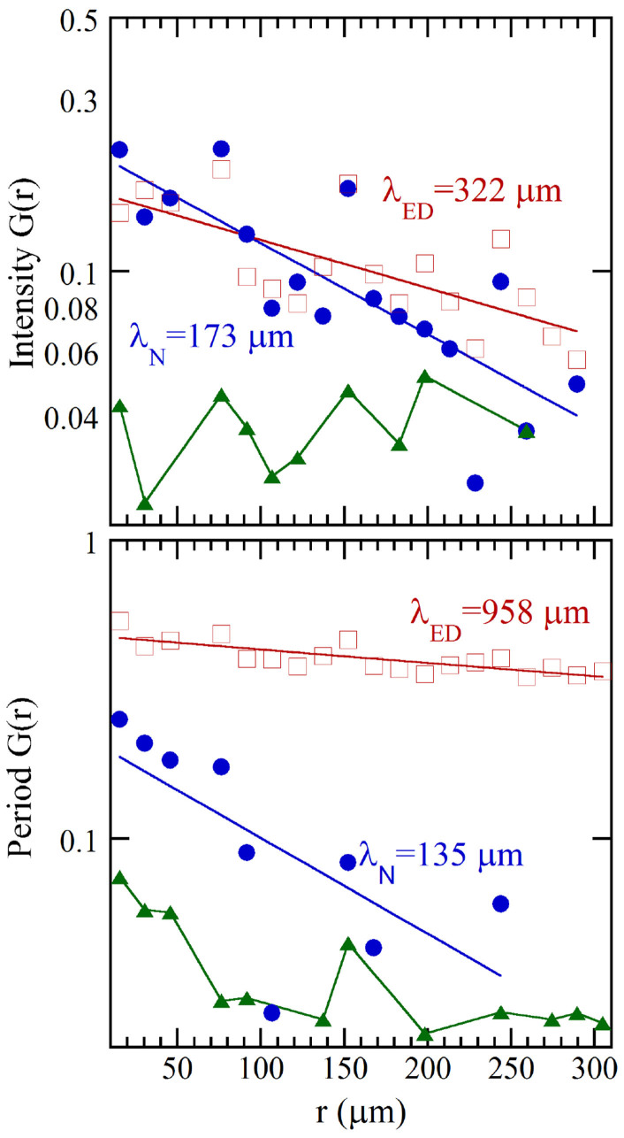
Degradation of the myelin sheath is a common pathology underlying demyelinating neurological diseases from Multiple Sclerosis to Leukodistrophies. Although large malformations of myelin ultrastructure in the advanced stages of Wallerian degradation is known, its subtle structural variations at early stages of demyelination remains poorly characterized. This is partly due to the lack of suitable and non-invasive experimental probes possessing sufficient resolution to detect the degradation. Here we report the feasibility of the application of an innovative non-invasive local structure experimental approach for imaging the changes of statistical structural fluctuations in the first stage of myelin degeneration. Scanning micro X-ray diffraction, using advances in synchrotron x-ray beam focusing, fast data collection, paired with spatial statistical analysis, has been used to unveil temporal changes in the myelin structure of dissected nerves following extraction of the Xenopus laevis sciatic nerve. The early myelin degeneration is a specific ordered compacted phase preceding the swollen myelin phase of Wallerian degradation. Our demonstration of the feasibility of the statistical analysis of SµXRD measurements using biological tissue paves the way for further structural investigations of degradation and death of neurons and other cells and tissues in diverse pathological states where nanoscale structural changes may be uncovered.
Figures
Similar articles
-
Comparison of CNS and PNS myelin proteins in the pathology of myelin disorders.J Neurol Sci. 2005 Feb 15;228(2):187-9. doi: 10.1016/j.jns.2004.10.005. Epub 2004 Dec 1. J Neurol Sci. 2005. PMID: 15694201 Review. No abstract available.
-
Demyelination can proceed independently of axonal degradation during Wallerian degeneration in wlds mice.Eur J Neurosci. 2011 Aug;34(4):531-7. doi: 10.1111/j.1460-9568.2011.07783.x. Epub 2011 Jul 12. Eur J Neurosci. 2011. PMID: 21749497
-
Targeting inflammatory demyelinating lesions to sites of Wallerian degeneration.Am J Pathol. 2007 Nov;171(5):1563-75. doi: 10.2353/ajpath.2007.070147. Epub 2007 Sep 6. Am J Pathol. 2007. PMID: 17823280 Free PMC article.
-
Differential regulation of myelin phagocytosis by macrophages/microglia, involvement of target myelin, Fc receptors and activation by intravenous immunoglobulins.J Neurosci Res. 2002 Jan 15;67(2):185-90. doi: 10.1002/jnr.10104. J Neurosci Res. 2002. PMID: 11782962
-
A unified cell biological perspective on axon-myelin injury.J Cell Biol. 2014 Aug 4;206(3):335-45. doi: 10.1083/jcb.201404154. J Cell Biol. 2014. PMID: 25092654 Free PMC article. Review.
Cited by
-
Lipid Order Degradation in Autoimmune Demyelination Probed by Polarized Coherent Raman Microscopy.Biophys J. 2017 Oct 3;113(7):1520-1530. doi: 10.1016/j.bpj.2017.07.033. Biophys J. 2017. PMID: 28978445 Free PMC article.
References
-
- Madura T. Pathophysiology of Peripheral Nerve Injury, in Basic Principles of Peripheral Nerve Disorders Edited by Dr. S. M. Rayegani, “InTech” Rijeka, Croatia, 278 pages. ISBN 978-953-51-0407-02 (2012).
-
- Valentijin L. J., Baas F., Wolterman R. A., Hoodgendijik J. E., van den Bosch N. H., Zorn I., Gabreëls-Festen A. W., de Visser M. & Bolhuis P. A. Identical point mutations of PMP-22 in Trembler-J mouse and Charcot-Marie-Tooth disease type 1A. Nat. Gen. 2, 288–291 (1992). - PubMed
-
- Dutta R. & Trapp B. D. Pathogenesis of axonal and neuronal damage in multiple sclerosis. Neurology 68, S22–S31 (2007). - PubMed
-
- McFarland H. F. & Martin R. Multiple sclerosis: a complicated picture of autoimmunity. Nat. Immunol. 8, 913–919 (2007). - PubMed
Publication types
MeSH terms
Grants and funding
LinkOut - more resources
Full Text Sources
Other Literature Sources
