. 2014 Jun 30;5(12):4426-37.
doi: 10.18632/oncotarget.2032.

Apelin promotes lymphangiogenesis and lymph node metastasis

Affiliations

Apelin promotes lymphangiogenesis and lymph node metastasis

Judit Berta et al. Oncotarget. .

Abstract

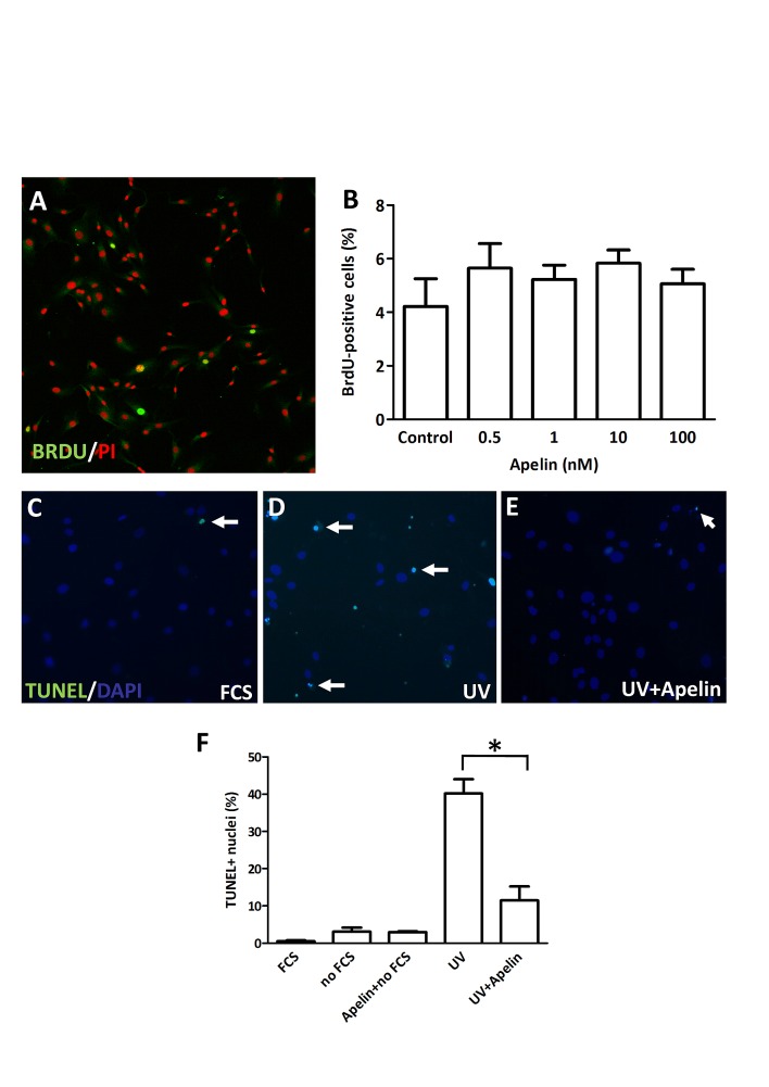
Whereas the role of the G-protein-coupled APJ receptor and its ligand, apelin, in angiogenesis has been well documented, the ability of the apelin/APJ system to induce lymphangiogenesis and lymphatic metastasis has been largely unexplored. To this end, we first show that APJ is expressed in lymphatic endothelial cells (LECs) and, moreover, that it responds to apelin by activating the apelinergic signaling cascade. We find that although apelin treatment does not influence the proliferation of LECs in vitro, it enhances their migration, protects them against UV irradiation-induced apoptosis, increases their spheroid numbers in 3D culture, stimulates their in vitro capillary-like tube formation and, furthermore, promotes the invasive growth of lymphatic microvessels in vivo in the matrigel plug assay. We also demonstrate that apelin overexpression in malignant cells is associated with accelerated in vivo tumor growth and with increased intratumoral lymphangiogenesis and lymph node metastasis. These results indicate that apelin induces lymphangiogenesis and, accordingly, plays an important role in lymphatic tumor progression. Our study does not only reveal apelin as a novel lymphangiogenic factor but might also open the door for the development of novel anticancer therapies targeting lymphangiogenesis.


Conflict of interest statement

The authors declare no potential conflicts of interest.

Figures

Figure 1
Figure 1. Expression and activation of APJ receptor in LECs
(A) In LECs, APJ mRNA was detected at a comparable level to that in HUVECs. (B), Immunofluorescent staining of APJ (green) in HUVECs and LECs. Nuclei were labeled with PI (red). (D), Time course of phosphorylation of Erk1/2, Akt and S6 following 1 μM apelin-13 treatment of LECs. (E), Quantification of activation by the ratio of the phosphorylated and total protein levels shows robust and significant activation of Erk1/2 and Akt. No significant activation of S6 was found upon apelin treatment. Asterisks designate significant differences (P<0.05). Columns represent mean of three experiments; bars, SEM.
Figure 2
Figure 2. Proliferation and apoptosis of LECs following apelin treatment
(A), LECs were cultured in serum-free medium and treated with apelin. After 96 hours, cells were pulsed with BrdU, fixed and stained with an anti-BrdU antibody (green) and PI (red). (B), No significant change was found in the ratio of BrdU-labeled nuclei upon apelin treatments. (C-E), Apoptosis induction with UV in LECs. Apoptotic cells are labeled with TUNEL (green). DAPI nuclei staining is blue. White arrows indicate apoptotic nuclei. (F), Robust apoptosis was induced by UV irradiation. Apelin treatment significantly reduced the ratio of UV induced apoptotic cells. Asterisks designate significant differences (P<0.05). In (B) and (F), columns represent mean of three experiments; bars, SD.
Figure 3
Figure 3. Effect of apelin on LEC migration
(A-B), Representative trajectories on the last phase contrast image of the path of LECs with (B) or without (A) apelin treatment. The color of the depicted trajectories refers to the elapsed time in the order from red–green–blue. (C), Average migrated distances of human LECs after incubation with apelin-13 and/or F13A (an APJ antagonist). (D), 12h average migrated distance of LECs after incubation with 100nM apelin-13 significantly increased cell migration and it was reduced in a dose dependent manner by the addition of F13A. Asterisks designate significant differences (P<0.05).
Figure 4
Figure 4. LEC spheroid formation in the presence of apelin
(A-B), LECs were plated in serum-free medium in non-adherent plates with apelin-13. After 96h incubation, all spheroids were photographed. (C-D), 1μM apelin-13 significantly increased the number of spheres without affecting their average diameter. Columns represent mean of three experiments; bars, SD; *, P<0.05 vs. control. Scale bar 100 μm
Figure 5
Figure 5. Tube formation capacity of LECs upon apelin treatment in vitro and in vivo.
(A-C), LECs (105) were plated on growth factor-reduced Matrigel-coated tissue culture plates in serum-free endothelial culture medium. 1 μM apelin-13 or 20 ng/ml bFGF was added to the medium. Capillary-like structures within the Matrigel layer were photographed after 18 hours. (D), Both apelin and bFGF significantly increased the average length of tubes (*, P<0.05 vs. control). Columns represent mean of three experiments; bars, SD; a.u., arbitrary unit (E), Matrigel containing PBS, apelin-13 (0.25 μg) or bFGF (0.25 μg) was injected subcutanously into mice. Both bFGF and apelin-13 significantly increased the density of LYVE-1 stained tubular structures in the sections of one-week old plugs. Columns represent means of three experiments; bars, SD; *, P<0.05 vs. control. LVDs are mean lymph vessel counts per square millimeter.
Figure 6
Figure 6. In vivo growth of apelin expressing tumor cells
(A), Overexpression of apelin through genetic manipulation significantly stimulated the in vivo growth of murine B16 melanoma cells in C57BL/6 mice. Growth curves of control vector (B16 Mock)- and apelin-transfected (B16 Apelin) cells. • and ■, means for 10 mice per group; bars, SD; *, P<0.05, versus control. (B-D), Apelin overexpression increases tumor-induced lymphangiogenesis in vivo. Frozen sections of 18-day-old control (B) and apelin-overexpressing (D) tumors were stained for the LEC marker LYVE-1 (red). Magnification, x200 (B, D). (C), LVDs (mean lymph vessel counts per square millimeter) of 18-day-old apelin-overexpressing or of control B16 tumors. Columns, means for ten mice per group; bars, SD; *, P<0.05 vs. control. (E-F), Apelin-overexpressing B16 cells also developed significantly larger tumors (lower row) when injected into the footpads of mice (*, P<0.05). Ipsilateral inguinal LN regions were carefully examined under a stereomicroscope, photographed and removed (G) (arrowhead shows pigmented B16 cells in the LN). (H), Histological demonstration of LN metastasis of B16 melanoma. Asterisk shows the colony of metastatic B16 cells in a LN. (I), The percentages of metastatic ipsilateral inguinal LNs (as evaluated by stereomicroscopy and histology) were 89% and 30% in the B16-Apelin and B16-Mock groups, respectively (*, P<0.05).

References

    1. Karpanen T, Alitalo K. Molecular biology and pathology of lymphangiogenesis. Annu Rev Pathol. 2008;3:367–397. - PubMed
    1. Saito Y, Nakagami H, Kaneda Y, Morishita R. Lymphedema and therapeutic lymphangiogenesis. Biomed Res Int. 2013;2013:804675. - PMC - PubMed
    1. Stacker SA, Williams SP, Karnezis T, Shayan R, Fox SB, Achen MG. Lymphangiogenesis and lymphatic vessel remodelling in cancer. Nat Rev Cancer. 2014;14(3):159–172. - PubMed
    1. Pitkin SL, Maguire JJ, Bonner TI, Davenport AP. International Union of Basic and Clinical Pharmacology. LXXIV. Apelin receptor nomenclature, distribution, pharmacology, and function. Pharmacol Rev. 2010;62(3):331–342. - PubMed
    1. Devic E, Rizzoti K, Bodin S, Knibiehler B, Audigier Y. Amino acid sequence and embryonic expression of msr/apj, the mouse homolog of Xenopus X-msr and human APJ. Mech Dev. 1999;84(1-2):199–203. - PubMed

Publication types