. 2015 Nov;25(11):4191-202.
doi: 10.1093/cercor/bhu142. Epub 2014 Jun 24.

Functional Brain Activity Relates to 0-3 and 3-8 Hz Force Oscillations in Essential Tremor

Affiliations

Functional Brain Activity Relates to 0-3 and 3-8 Hz Force Oscillations in Essential Tremor

Kristina A Neely et al. Cereb Cortex. 2015 Nov.

Abstract

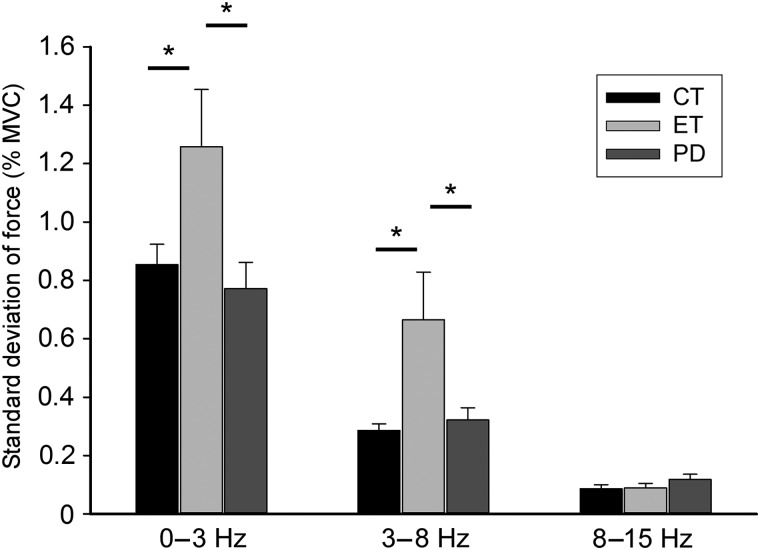
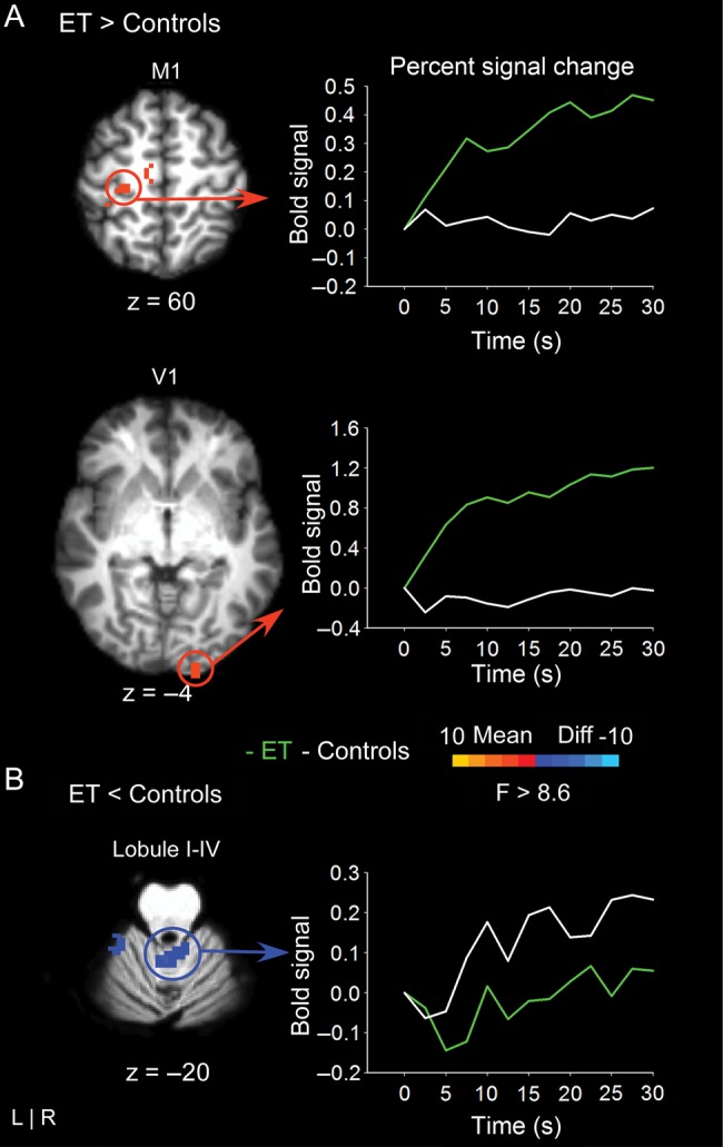
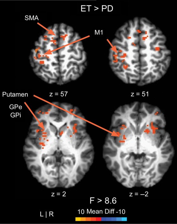
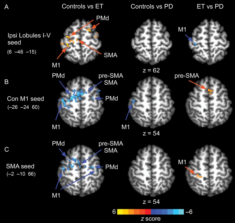
It is well-established that during goal-directed motor tasks, patients with essential tremor have increased oscillations in the 0-3 and 3-8 Hz bands. It remains unclear if these increased oscillations relate to activity in specific brain regions. This study used task-based functional magnetic resonance imaging to compare the brain activity associated with oscillations in grip force output between patients with essential tremor, patients with Parkinson's disease who had clinically evident tremor, and healthy controls. The findings demonstrate that patients with essential tremor have increased brain activity in the motor cortex and supplementary motor area compared with controls, and this activity correlated positively with 3-8 Hz force oscillations. Brain activity in cerebellar lobules I-V was reduced in essential tremor compared with controls and correlated negatively with 0-3 Hz force oscillations. Widespread differences in brain activity were observed between essential tremor and Parkinson's disease. Using functional connectivity analyses during the task evidenced reduced cerebellar-cortical functional connectivity in patients with essential tremor compared with controls and Parkinson's disease. This study provides new evidence that in essential tremor 3-8 Hz force oscillations relate to hyperactivity in motor cortex, 0-3 Hz force oscillations relate to the hypoactivity in the cerebellum, and cerebellar-cortical functional connectivity is impaired.

Keywords: Parkinson's disease; cerebellum; essential tremor; fMRI; grip force; motor control.


Figures

Figure 1.
Figure 1.
Methods for the precision grip task. (A) Modified precision grip shown with custom-built force transducer. (B) The visual display consists of 2 bars: a moveable red/green force bar and a (stationary) white target bar. The red bar turned green allowing it to be moved up and down in direct accordance to the amount of force applied to the transducer. The location of the white bar indicated the target amplitude. A green force bar indicated “go” and a red force bar indicated “rest”. (C) A representative raw force trace from a healthy participant. Participants completed 10, 2-s force pulses separated by 1 s of rest for 30 s.
Figure 2.
Figure 2.
The standard deviation (%MVC) of force output for essential tremor patients, Parkinson's disease patients, and controls in each filtered dataset. We show 0–3, 3–8, and 8–15 Hz bands. Error bars are one standard error from the mean. Significant findings are noted with a star.
Figure 3.
Figure 3.
The results for the voxelwise analysis comparing essential tremor and controls are shown in 4 slices of the cortex. (A) At left, regions of hyperactivation are shown in left primary motor cortex (M1) and right primary visual cortex (V1). The corresponding line graph to the right shows that percent signal change is higher for essential tremor patients compared with controls across the time-series. The time-point “0” designates the onset of the 30-s force interval. (B) At left, regions of hypoactivation within the cerebellum are shown. The corresponding line graph to the right shows that percent signal change is lower for essential tremor patients compared with controls across the time-series. The time-point “0” designates the onset of the 30-s force interval.
Figure 4.
Figure 4.
The results for the voxelwise analysis comparing essential tremor and Parkinson's disease patients are shown in 4 slices of the cortex and subcortex. All voxels with differences between groups showed higher BOLD signal for essential tremor patients compared with Parkinson's disease patients.
Figure 5.
Figure 5.
Functional connectivity for a seed in right lobule I–V (A), left M1 (B), and SMA (C). Comparisons of functional connectivity were made between controls versus essential tremor, controls versus Parkinson's disease, and essential tremor versus Parkinson's disease.

Similar articles

Cited by

References

    1. Axelrad JE, Louis ED, Honig LS, Flores I, Ross GW, Pahwa R, Lyons KE, Faust PL, Vonsattel JP. 2008. Reduced Purkinje cell number in essential tremor: a postmortem study. Arch Neurol. 65:101–107. - PMC - PubMed
    1. Baarbe J, Yielder P, Daligadu J, Behbahani H, Haavik H, Murphy B. 2014. A novel protocol to investigate motor training-induced plasticity and sensorimotor integration in the cerebellum and motor cortex. J Neurophysiol. 111:715–721. - PubMed
    1. Bell EC, Willson MC, Wilman AH, Dave S, Silverstone PH. 2006. Males and females differ in brain activation during cognitive tasks. Neuroimage. 30:529–538. - PubMed
    1. Bernard JA, Peltier SJ, Benson BL, Wiggins JL, Jaeggi SM, Buschkuehl M, Jonides J, Monk CS, Seidler RD. 2014. Dissociable functional networks of the human dentate nucleus. Cereb Cortex. 24:2151–2159. - PMC - PubMed
    1. Bernard JA, Seidler RD, Hassevoort KM, Benson BL, Welsh RC, Wiggins JL, Jaeggi SM, Buschkuehl M, Monk CS, Jonides J, et al. 2012. Resting state cortico-cerebellar functional connectivity networks: a comparison of anatomical and self-organizing map approaches. Front Neuroanat. 6:31. - PMC - PubMed

Publication types