. 2014 Sep 1;20(17):4636-46.
doi: 10.1158/1078-0432.CCR-14-0305. Epub 2014 Jun 24.

miR-409-3p/-5p promotes tumorigenesis, epithelial-to-mesenchymal transition, and bone metastasis of human prostate cancer

Affiliations

miR-409-3p/-5p promotes tumorigenesis, epithelial-to-mesenchymal transition, and bone metastasis of human prostate cancer

Sajni Josson et al. Clin Cancer Res. .

Abstract

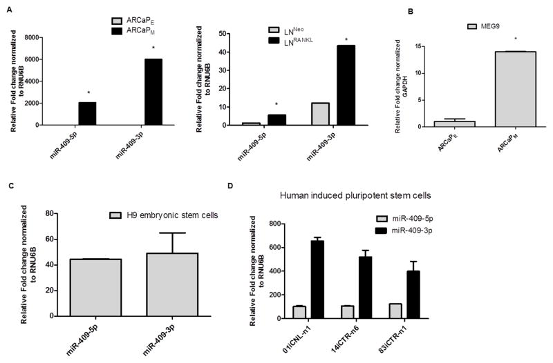
Purpose: miR-409-3p/-5p is a miRNA expressed by embryonic stem cells, and its role in cancer biology and metastasis is unknown. Our pilot studies demonstrated elevated miR-409-3p/-5p expression in human prostate cancer bone metastatic cell lines; therefore, we defined the biologic impact of manipulation of miR-409-3p/-5p on prostate cancer progression and correlated the levels of its expression with clinical human prostate cancer bone metastatic specimens.

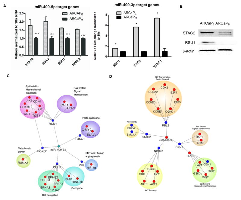
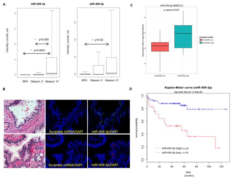
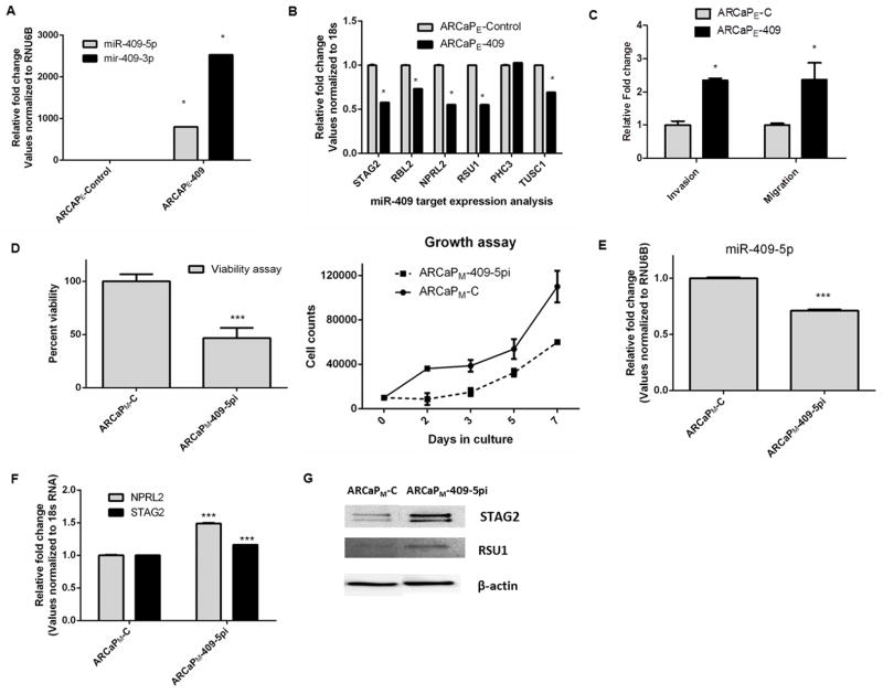
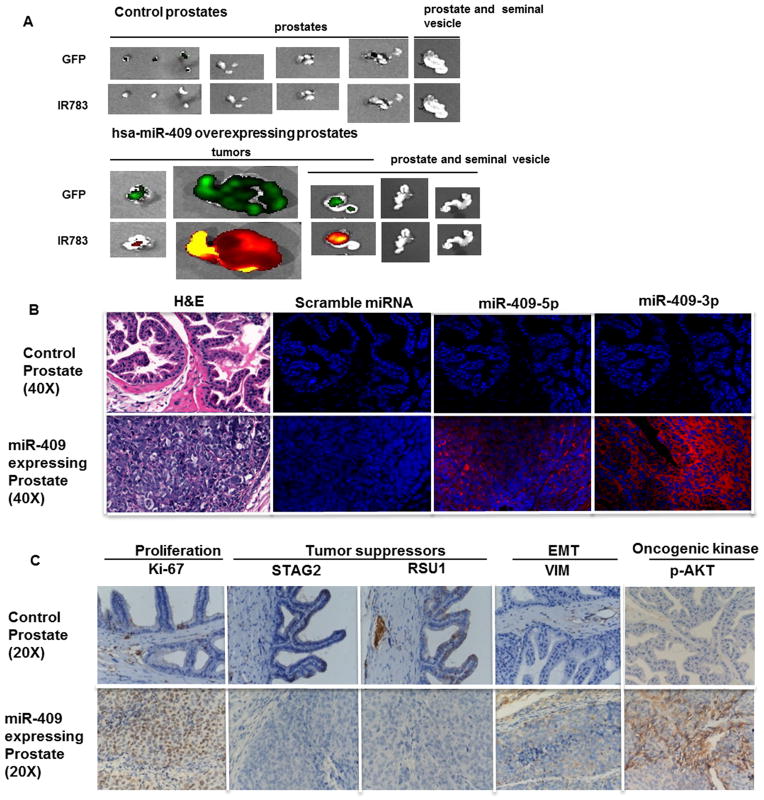
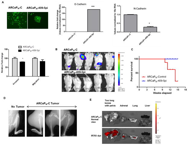
Experimental design: miRNA profiling of a prostate cancer bone metastatic epithelial-to-mesenchymal transition (EMT) cell line model was performed. A Gleason score human tissue array was probed for validation of specific miRNAs. In addition, genetic manipulation of miR-409-3p/-5p was performed to determine its role in tumor growth, EMT, and bone metastasis in mouse models.

Results: Elevated expression of miR-409-3p/-5p was observed in bone metastatic prostate cancer cell lines and human prostate cancer tissues with higher Gleason scores. Elevated miR-409-3p expression levels correlated with progression-free survival of patients with prostate cancer. Orthotopic delivery of miR-409-3p/-5p in the murine prostate gland induced tumors where the tumors expressed EMT and stemness markers. Intracardiac inoculation (to mimic systemic dissemination) of miR-409-5p inhibitor-treated bone metastatic ARCaPM prostate cancer cells in mice led to decreased bone metastasis and increased survival compared with control vehicle-treated cells.

Conclusion: miR-409-3p/-5p plays an important role in prostate cancer biology by facilitating tumor growth, EMT, and bone metastasis. This finding bears particular translational importance as miR-409-3p/-5p appears to be an attractive biomarker and/or possibly a therapeutic target to treat bone metastatic prostate cancer.


Figures

Figure 1
Figure 1
MicroRNA miR-409-3p/-5p in the imprinted DLK1-DIO3 cluster is overexpressed in bone metastatic EMT models of human PCa. A, miR-409-3p/-5p in bone metastatic PCa models (mesenchymal cells ARCaPM compared to ARCaPE) and (LNCaPNeo verses LNCaPRANKL PCa cells). All miRNA and RNA analysis was performed by qRT-PCR analysis. B, mRNA levels of MEG9 of ARCaPE and ARCaPM PCa cells. C, miR-409-3p/-5p expression in H9 embryonic stem cells and, D, iPSCs. *: p<0.05 were considered to be statistically significant by t-test.
Figure 2
Figure 2
miR-409 inhibits tumor suppressor genes in prostate cancer. A, mRNA targets of miR-409-5p: STAG2, RBL2, RSU1 and NPRL2 and mRNA targets of miR-409-3p: RSU1, PHC3 and TUSC1, assayed by triplicate wells in qRT-PCR of ARCaPE and ARCaPM cells. The representative RT-PCR is shown. The experiment was repeated twice. B, Western analysis of STAG2 and RSU1 in ARCaPE and ARCaPM PCa cells. C and D, Cytoscape images of miR-409-3p and miR-409-5p signaling pathways. *: p<0.05 were considered to be statistically significant by t-test.
Figure 3
Figure 3
Human prostatic tissues with higher Gleason score and prostate cancer bone metastasis tissues express elevated levels of miR-409. A, Quantitative analysis of miR-409-3p and miR-409-5p expression in tumor tissues with Gleason grade. B, Representative image of miR-409-3p (green) and miR-409-5p (red) expression in tumor tissues and H&E staining (40x). The tissue array consisted of BPH (N=14), Gleason 6 (N=26) and Gleason ≥ 7 (N=35), data analyzed by Kruskal-Wallis one way analysis of variance- Tukey method. C, miR-409-3p expression in Gleason_high (N=29) and Gleason_low (N=78) based on MSKCC dataset. D, Kaplan-Meier disease free survival (DFS) curves for the prostate cancer patients, based on miR-409-3p expression in the MSKCC dataset. The y-axis is disease free survival probability, and the x-axis is survival in months. Blue line represents the DFS of patients with miR-409-3p lower than the median of the normal individuals (n=78). Red line represents the DFS of patients with miR-409-3p higher than the median of the normal individuals (n=29). Data was analyzed using log-rank test (p=4.3e-05). *: p<0.05 were considered to be statistically significant.
Figure 4
Figure 4
Ectopic expression of miR-409 leads to increased invasiveness and aggressiveness of prostate cancer cells and conversely inhibition of miR-409 results in increased cell death in PCa cells. A, miR-409-5p and -3p expression by qRT-PCR in ARCaPE-C and ARCaPE-409 expressing PCa cells. B, RNA expression of miR-409-5p/-3p targets in ARCaPE-C and ARCaPE-409 expressing PCa cells assayed by real time PCR. (miR-409-5p mRNA targets: STAG2, RBL2, NPRL2 and RSU1and miR-409-3p mRNA targets: RSU1, PHC3 and TUSC1). C, Invasion and migration assay of in ARCaPE-C and ARCaPE-409 expressing PCa cells. D, Cell viability in ARCaPM PCa cells in response to a miR-409-5p inhibitor. Growth curve of ARCaPM-C and ARCaPM-409-5pi PCa cells. E, Expression of miR-409-5p assayed by qRT- PCR in ARCaPM-C control PCa and ARCaPM-409-5pi (miR-409-5p inhibitor transfected cells). F, RNA expression of miR-409-5p targets in ARCaPM-C control and ARCaPM-409-5pi cells assayed by qRT- PCR. (miR-409-5p mRNA targets: NPRL2 and STAG2). G, Protein expression of STAG2 and RSU1 in ARCaPM-C cells and ARCaPM-409-5pi cells. *: p<0.05 were considered to be statistically significant by t-test.
Figure 5
Figure 5
Ectopic expression of miR-409 in the prostate gland transforms normal prostate epithelia, promotes tumorigenecity, EMT and stemness in vivo. A, Comparison of normal prostate and miR-409 expressing prostates. Top row represents green fluorescence for cells containing control GFP plasmid or miR-409 GFP expressing plasmid. Bottom panel represent tumor specific NIR dye (IR783) uptake in control or miR-409 expressing prostates. B, H&E staining of normal control prostate and adenocarcinoma lesions of miR-409 overexpressing prostates (40x), followed by miRNA detection of scramble miRNA and miR-409-5p/-3p of control and miR-409 expressing tissues by ISH and QD detection (40x). C, IHC staining of Ki-67, STAG2, RSU1, vimentin and p-AKT in control prostate and miR-409 expressing prostate tissues (20x).
Figure 6
Figure 6
Inhibition of miR-409-5p results in decreased bone metastasis in PCa in vivo. A, Morphological EMT changes in miR-409-5p inhibited ARCaPM cells; magnification 10x. RNA expression assayed by qRT-PCR of EMT markers, E-cadherin and N-cadherin. Migration and invasion assay of ARCaPM-C and ARCaPM-409-5pi PCa cells (n=3). B, Metastatic lesions observed by luciferase imaging of tumors of ARCaPM–C cells and ARCaPM-409-5pi cells in SCID/Beige mice following intra-cardial injections (N=5). C, Kaplan Meier survival curve of mice injected with ARCaPM–C cells and ARCaPM-409-5pi cells mice. D, X-ray image of metastatic bone lesion from ARCaPM–C bone tumors. E, Tumor dye (IR-783 dye) uptake by ARCaPM–C metastatic tumors from a representative mouse.

References

    1. Ma L, Teruya-Feldstein J, Weinberg RA. Tumour invasion and metastasis initiated by microRNA-10b in breast cancer. Nature. 2007;449:682–8. - PubMed
    1. Tavazoie SF, Alarcon C, Oskarsson T, Padua D, Wang Q, Bos PD, et al. Endogenous human microRNAs that suppress breast cancer metastasis. Nature. 2008;451:147–52. - PMC - PubMed
    1. Zhang Y, Yang P, Sun T, Li D, Xu X, Rui Y, et al. miR-126 and miR-126* repress recruitment of mesenchymal stem cells and inflammatory monocytes to inhibit breast cancer metastasis. Nat Cell Biol. 2013;15:284–94. - PMC - PubMed
    1. Korpal M, Ell BJ, Buffa FM, Ibrahim T, Blanco MA, Celia-Terrassa T, et al. Direct targeting of Sec23a by miR-200s influences cancer cell secretome and promotes metastatic colonization. Nat Med. 2011;17:1101–8. - PMC - PubMed
    1. Chou J, Lin JH, Brenot A, Kim JW, Provot S, Werb Z. GATA3 suppresses metastasis and modulates the tumour microenvironment by regulating microRNA-29b expression. Nat Cell Biol. 2013;15:201–13. - PMC - PubMed

Publication types