

Expression of IL-18, IL-18 binding protein, and IL-18 receptor by normal and cancerous human ovarian tissues: possible implication of IL-18 in the pathogenesis of ovarian carcinoma
- PMID: 24963217
- PMCID: PMC4052106
- DOI: 10.1155/2014/914954
Expression of IL-18, IL-18 binding protein, and IL-18 receptor by normal and cancerous human ovarian tissues: possible implication of IL-18 in the pathogenesis of ovarian carcinoma
Abstract
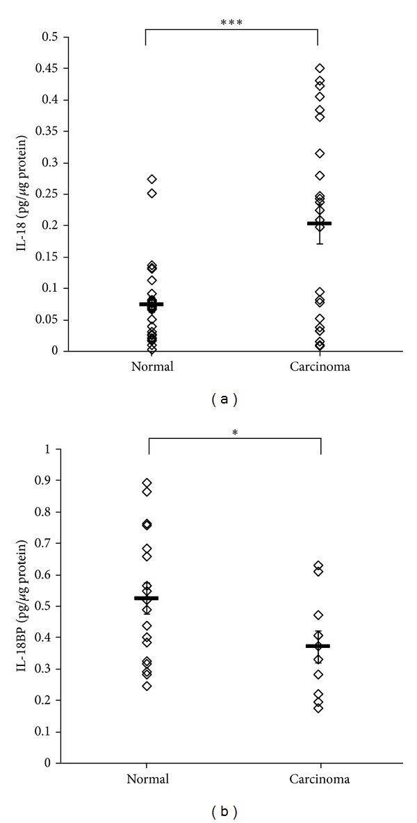
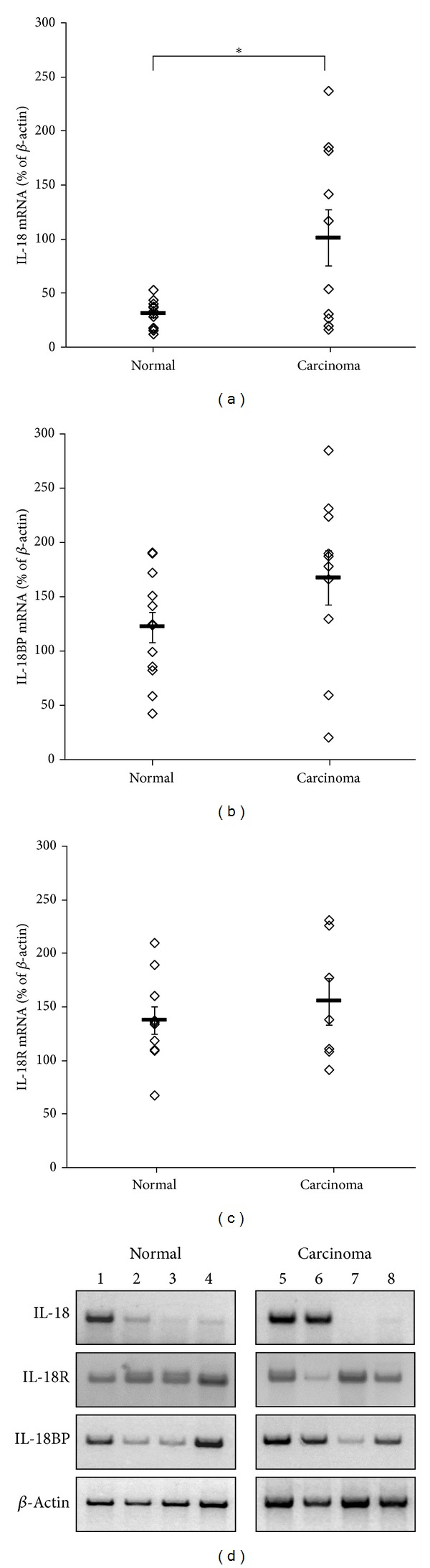
Proinflammatory cytokine IL-18 has been shown to be elevated in the sera of ovarian carcinoma patients. The aim of the study was to examine the levels and cellular origin of IL-18, IL-18 binding protein, and IL-18 receptor in normal and cancerous ovarian tissues. Ovarian tissue samples were examined by immunohistochemical staining for IL-18, IL-18BP, and IL-18R and mRNA of these cytokines was analyzed with semiquantitative PT-PCR. IL-18 levels were significantly higher in cancerous ovarian tissues (P = 0.0007), IL-18BP levels were significantly higher in normal ovarian tissues (P = 0.04), and the ratio of IL-18/IL-18BP was significantly higher in cancerous ovarian tissues (P = 0.036). Cancerous ovarian tissues expressed significantly higher IL-18 mRNA levels (P = 0.025), while there was no difference in the expression of IL-18BP mRNA and IL-18R mRNA between cancerous and normal ovarian tissues. IL-18 and IL-18BP were expressed dominantly in the epithelial cells of both cancerous and normal ovarian tissues, while IL-18R was expressed dominantly in the epithelial cells of cancerous ovarian tissues but expressed similarly in the epithelial and stromal cells of normal cancerous tissues. This study indicates a possible role of IL-18, IL-18BP, and IL-18R in the pathogenesis of epithelial ovarian carcinoma.
Figures

Similar articles
-
Expression of IL-10 in human normal and cancerous ovarian tissues and cells.Eur Cytokine Netw. 2010 Jun;21(2):122-8. doi: 10.1684/ecn.2010.0188. Epub 2010 Apr 30. Eur Cytokine Netw. 2010. PMID: 20430716
-
The IL-18 antagonist IL-18-binding protein is produced in the human ovarian cancer microenvironment.Clin Cancer Res. 2013 Sep 1;19(17):4611-20. doi: 10.1158/1078-0432.CCR-13-0568. Epub 2013 Jul 19. Clin Cancer Res. 2013. PMID: 23873689
-
Interleukin-18 system messenger RNA and protein expression in human endometrium during the menstrual cycle.Fertil Steril. 2006 Oct;86(4):905-13. doi: 10.1016/j.fertnstert.2006.02.122. Fertil Steril. 2006. PMID: 17027359
-
Interleukin 18 and interleukin 18 binding protein: possible role in immunosuppression of chronic renal failure.Blood Purif. 2003;21(3):258-70. doi: 10.1159/000070699. Blood Purif. 2003. PMID: 12784053 Review.
-
IL-18 and IL-18BP: A Unique Dyad in Health and Disease.Int J Mol Sci. 2024 Dec 17;25(24):13505. doi: 10.3390/ijms252413505. Int J Mol Sci. 2024. PMID: 39769266 Free PMC article. Review.
Cited by
-
New immunological potential markers for triple negative breast cancer: IL18R1, CD53, TRIM, Jaw1, LTB, PTPRCAP.Discov Oncol. 2021 Mar 10;12(1):6. doi: 10.1007/s12672-021-00401-0. Discov Oncol. 2021. PMID: 35201443 Free PMC article.
-
Interleukin‑18 binding protein: Biological properties and roles in human and animal immune regulation (Review).Biomed Rep. 2024 Apr 9;20(6):87. doi: 10.3892/br.2024.1775. eCollection 2024 Jun. Biomed Rep. 2024. PMID: 38665423 Free PMC article. Review.
-
The Possible Role of Interleukin (IL)-18 and Nitrous Oxide and Their Relation to Oxidative Stress in the Development and Progression of Breast Cancer.Asian Pac J Cancer Prev. 2019 Sep 1;20(9):2659-2665. doi: 10.31557/APJCP.2019.20.9.2659. Asian Pac J Cancer Prev. 2019. PMID: 31554361 Free PMC article.
-
Alpha Mangostin and Cisplatin as Modulators of Exosomal Interaction of Ovarian Cancer Cell with Fibroblasts.Int J Mol Sci. 2022 Aug 10;23(16):8913. doi: 10.3390/ijms23168913. Int J Mol Sci. 2022. PMID: 36012171 Free PMC article.
-
The Dual Role of STAT1 in Ovarian Cancer: Insight Into Molecular Mechanisms and Application Potentials.Front Cell Dev Biol. 2021 Mar 23;9:636595. doi: 10.3389/fcell.2021.636595. eCollection 2021. Front Cell Dev Biol. 2021. PMID: 33834023 Free PMC article. Review.
References
-
- Siegel R, Naishadham D, Jemal A. Cancer statistics, 2012. CA: A Cancer Journal for Clinicians. 2012;62(1):10–29. - PubMed
-
- Apte RN, Krelin Y, Song X, et al. Effects of micro-environment- and malignant cell-derived interleukin-1 in carcinogenesis, tumour invasiveness and tumour-host interactions. European Journal of Cancer. 2006;42(6):751–759. - PubMed
-
- Huleihel M, Maymon E, Piura B, et al. Distinct patterns of expression of interleukin-1 α and β by normal and cancerous human ovarian tissues. European Cytokine Network. 1997;8(2):179–187. - PubMed
-
- Glezerman M, Mazot M, Maymon E, et al. Tumor necrosis factor-alpha and interleukin-6 are differently expressed by fresh human cancerous ovarian tissue and primary cell lines. European Cytokine Network. 1998;9(2):171–179. - PubMed
-
- Rabinovich A, Medina L, Piura B, Segal S, Huleihel M. Regulation of ovarian carcinoma SKOV-3 cell proliferation and secretion of MMPs by autocrine IL-6. Anticancer Research. 2007;27(1A):267–272. - PubMed
MeSH terms
Substances
LinkOut - more resources
Full Text Sources
Other Literature Sources
Medical
Miscellaneous
