

Investigation of antimicrobial activity of photothermal therapeutic gold/copper sulfide core/shell nanoparticles to bacterial spores and cells
- PMID: 24963345
- PMCID: PMC4068869
- DOI: 10.1186/1754-1611-8-11
Investigation of antimicrobial activity of photothermal therapeutic gold/copper sulfide core/shell nanoparticles to bacterial spores and cells
Abstract
Background: Au/CuS core/shell nanoparticles (NPs) were designed as a new type of transducer agent for photothermal therapy (PTT), with attractive features of easy preparation, low cost and small size for targeting. This paper studied for the first time the intrinsic antimicrobial activity of Au/CuS NPs to B. anthracis spores and cells in addition to its PTT effect.
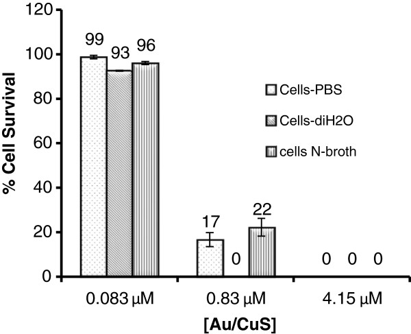
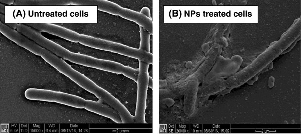
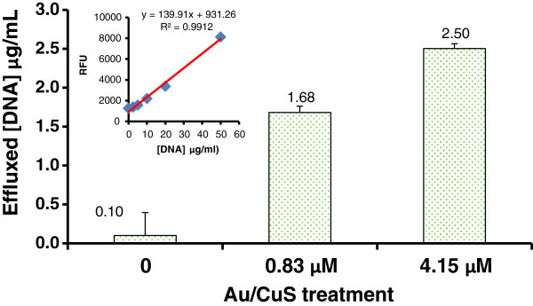
Results: It was found that Au/CuS NPs were highly efficient in inactivating B. anthracis cells, but not effective to the spores. Treatment with NPs at ~0.83 μM for 30 min achieved a 7 log reduction in viable cells. The antimicrobial effect was both NPs concentration and treatment time dependent. SEM imaging and the efflux of DNA test demonstrated the damage of cell membrane after NPs treatment, yet further research is necessary to fully understand the precise inactivation mechanism.
Conclusions: The Au/CuS NPs had strong antimicrobial activity to B. anthracis cells, which showed a great potential to be an effective antimicrobial agent to bacterial cells.
Keywords: Antimicrobial; Bacillus anthracis; Bioterrorism; Nanoparticles.
Figures
References
-
- Elsaka SE, Hamouda IM, Swain MV. Titanium dioxide nanoparticles addition to a conventional glass-ionomer restorative: influence on physical and antibacterial properties. J Dent. 2011;8:589–598. - PubMed
-
- Sondi I, Salopek-Sondi B. Silver nanoparticles as antimicrobial agent: a case study on E. coli as a model for Gram-negative bacteria. J Colloid Interface Sci. 2004;8:177–182. - PubMed
-
- Stoimenov PK, Klinger RL, Marchin GL, Klabunde KJ. Metal Oxide Nanoparticles as Bactericidal Agents. Langmuir. 2002;8:6679–6686.
-
- Zawrah M, El-Moez SA, Center D. Antimicrobial activities of gold nanoparticles against major foodborne pathogens. Life Sci J. 2011;8:37–44.
-
- Lin J, Zhang H, Chen Z, Zheng Y. Penetration of lipid membranes by gold nanoparticles: insights into cellular uptake, cytotoxicity, and their relationship. ACS Nano. 1021;8:5421–5429. - PubMed
LinkOut - more resources
Full Text Sources
Other Literature Sources
