

MRI brain findings in ephedrone encephalopathy associated with manganese abuse: Single-center perspective
- PMID: 24963359
- PMCID: PMC4067420
- DOI: 10.12659/PJR.889690
MRI brain findings in ephedrone encephalopathy associated with manganese abuse: Single-center perspective
Abstract
Background: Manganese (Mn) is a well-known toxic agent causing symptoms of parkinsonism in employees of certain branches of industry. Home production of a psychostimulant ephedrone (methcathinone), involving the use of potassium permanganate, became a new cause of intoxications in Poland.
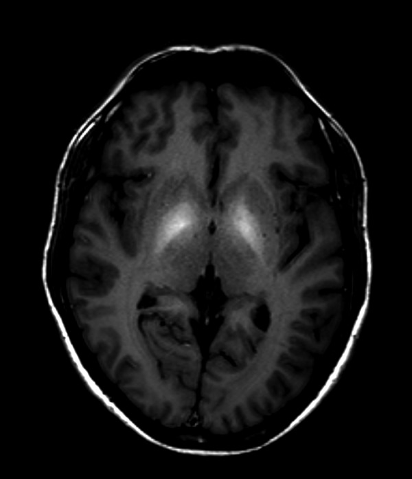
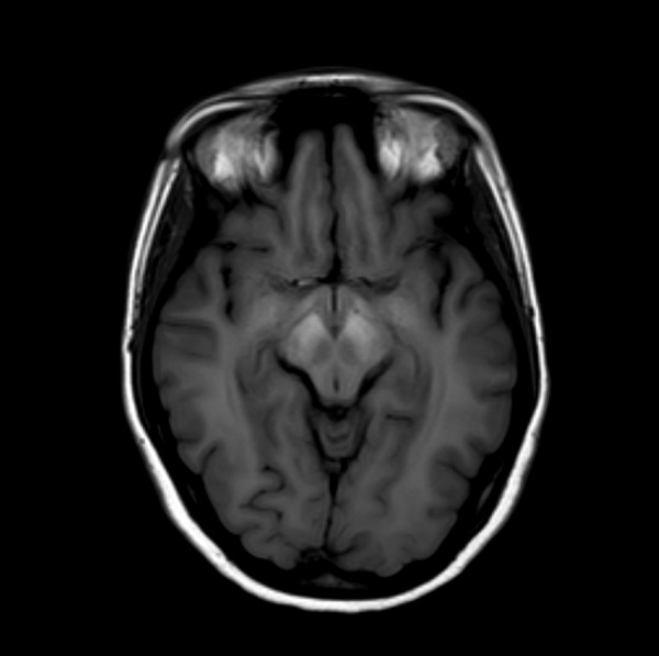
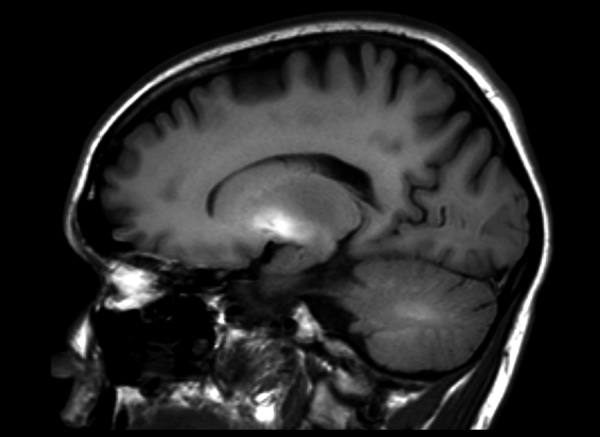
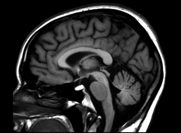
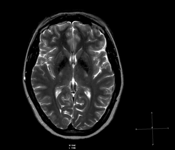
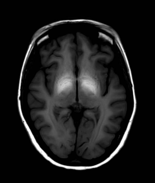
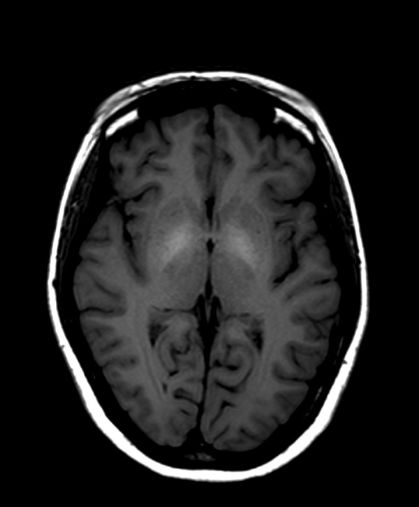
Case report: This article presents clinical symptoms, initial brain MRI findings and characteristics of changes observed in follow-up examinations in 4 patients with manganese intoxication associated with intravenous administration of ephedrone. All patients in our case series presented symptoms of parkinsonism. T1-WI MRI revealed high intensity signal in globi pallidi in all patients; hyperintense lesions in midbrain were observed in three patients, while lesions located in cerebellar hemispheres and pituitary gland in just one patient. The reduction of signal intensity in the affected brain structures was observed in follow-up studies, with no significant improvement in clinical symptoms.
Conclusions: Brain MRI is helpful in the assessment of distribution as well as dynamics of changes in ephedrone encephalopathy. Regression of signal intensity changes visible in brain MRI is not associated with clinical condition improvement. Although brain MRI findings are not characteristic for ephedrone encephalopathy, they may contribute to diagnosing this condition.
Keywords: Brain Diseases; Manganese Compounds; Metabolic - ultrasonography.
Figures
References
-
- Schmidt T, Dalubaeva D. Anniversary Collection: Diagnostic and Treatment of Neurological Diseases. W. Medicine Moscow; Russia: 1990. Neurological complications of ephedrone drug abuse (ephedrone encephalopathy) pp. 183–86.
-
- Sanotsky Y, Lesyk R, Fedoryshyn L, et al. Manganic encephalopathy due to “ephedrone” abuse”. Mov Disord. 2007;22(9):1337–43. - PubMed
-
- de Bie RM, Gladstone RM, Strafella AP, et al. Manganese-induced Parkinsonism associated with methcathinone (Ephedrone) abuse. Arch Neurol. 2007;64(6):886–89. - PubMed
Publication types
LinkOut - more resources
Full Text Sources
Other Literature Sources