. 2014:2014:812678.
doi: 10.1155/2014/812678. Epub 2014 May 20.

Fibroblast-like synoviocytes induce calcium mineral formation and deposition

Affiliations

Fibroblast-like synoviocytes induce calcium mineral formation and deposition

Yubo Sun et al. Arthritis. 2014.

Abstract

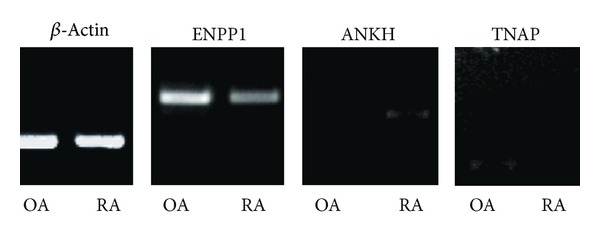
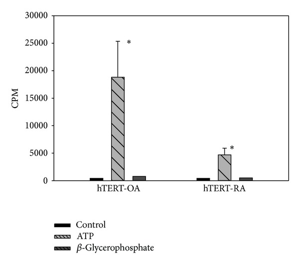
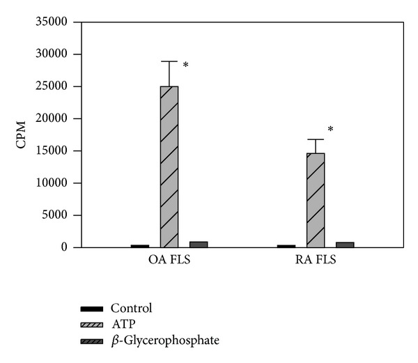
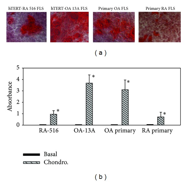
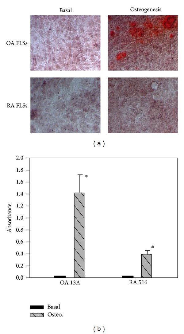
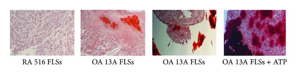
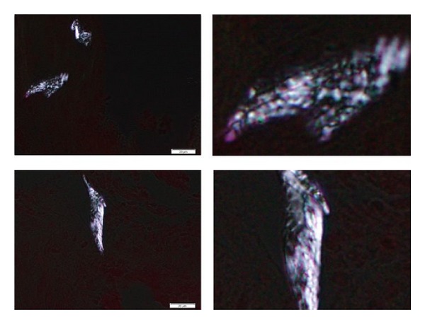
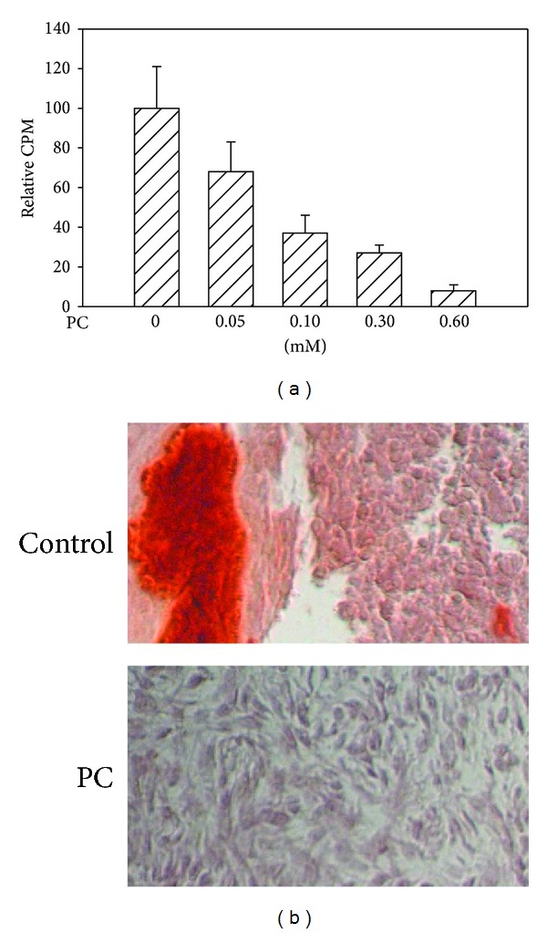
Calcium crystals are present in the synovial fluid of 65%-100% patients with osteoarthritis (OA) and 20%-39% patients with rheumatoid arthritis (RA). This study sought to investigate the role of fibroblast-like synoviocytes (FLSs) in calcium mineral formation. We found that numerous genes classified in the biomineral formation process, including bone gamma-carboxyglutamate (gla) protein/osteocalcin, runt-related transcription factor 2, ankylosis progressive homolog, and parathyroid hormone-like hormone, were differentially expressed in the OA and RA FLSs. Calcium deposits were detected in FLSs cultured in regular medium in the presence of ATP and FLSs cultured in chondrogenesis medium in the absence of ATP. More calcium minerals were deposited in the cultures of OA FLSs than in the cultures of RA FLSs. Examination of the micromass stained with nonaqueous alcoholic eosin indicated the presence of birefringent crystals. Phosphocitrate inhibited the OA FLSs-mediated calcium mineral deposition. These findings together suggest that OA FLSs are not passive bystanders but are active players in the pathological calcification process occurring in OA and that potential calcification stimuli for OA FLSs-mediated calcium deposition include ATP and certain unidentified differentiation-inducing factor(s). The OA FLSs-mediated pathological calcification process is a valid target for the development of disease-modifying drug for OA therapy.


Figures

Figure 1
Figure 1
Semiquantitative RT-PCR. RNA samples were extracted from hTERT-OA 13A FLSs and hTERT-RA 516 FLSs. The messenger levels of ENPP1, ANKH, and TNAP were determined by RT-PCR. β-Actin, 30 cycles; ENPP1, 35 cycles; ANKH, 40 cycles; and TNAP, 35 cycles.
Figure 2
Figure 2
ATP-induced calcium mineral formation and deposition in monolayer culture. Left bar group: untreated hTERT-OA 13A FLSs, ATP-treated hTERT-OA 13A FLSs, and beta-glycerophosphate treated hTERT-OA 13A FLSs (from left to right). Right bar group: untreated hTERT-RA 516 FLSs, ATP-treated hTERT-RA 516 FLSs, and beta-glycerophosphate treated hTERT-RA 516 FLSs (from left to right). Counts per minute (CPM) were normalized against total protein levels.
Figure 3
Figure 3
ATP-induced calcium mineral formation and deposition in monolayer culture of primary FLSs. Left panel: untreated OA FLSs, ATP-treated OA FLSs, and beta-glycerophosphate treated OA FLSs. Right panel: untreated RA FLSs, ATP-treated RA FLSs, and beta-glycerophosphate treated RA FLSs. Counts per minute (CPM) were normalized against total protein levels.
Figure 4
Figure 4
Alizarin red staining and reading. (a) Alizarin red staining of hTERT-RA 516 FLSs, hTERT-OA 13A FLSs, primary OA FLSs, and primary RA FLSs cultured in STEMPro chondrogenesis differentiation medium. (b) Absorbance of alizarin red extract from the monolayer cultures of hTERT-RA 516 FLSs, hTERT-OA 13A FLSs, primary OA FLSs, and primary RA FLSs cultured in the basal medium and STEMPro chondrogenesis differentiation medium.
Figure 5
Figure 5
Alizarin red staining and analysis.(a) Alizarin red staining of hTERT-OA 13A FLSs and hTERT-RA 516 FLSs cultured in basal medium and STEMPro osteogenesis differentiation medium. (b)Absorbance of alizarin red extract from the monolayer cultures of hTERT-OA 13A FLSs and hTERT-RA 516 FLSs cultured in basal medium and STEMPro osteogenesis differentiation medium.
Figure 6
Figure 6
Alizarin red staining. Micromass of hTERT-RA 516 FLSs cultured in the absence of ATP. There were no calcium deposits detected (first photo from the left). Micromass of hTERT-OA 13A FLSs cultured in the absence of ATP. Small amounts of calcium deposits were detected within the micromass (second photo from the left). Micromass of hTERT-OA 13A FLSs cultured in the absence of ATP. Large amounts of calcium deposits were detected in the fragments of the micromass, but not within the micromass (third photo from the left). Micromass of hTERT-OA 13A FLSs cultured in the presence of ATP. Large amounts of calcium deposits were detected within the micromass (far right photo).
Figure 7
Figure 7
Nonaqueous alcoholic eosin staining. Birefringent crystals in the micromasses of hTERT-OA 13A FLSs were observed using polarizing light microscope (left photos). Enlarged images of the respective birefringent crystals are shown in the corresponding right hand images.
Figure 8
Figure 8
Inhibition of calcium mineral formation and deposition. (a) PC inhibited ATP-induced calcium mineral formation and deposition in the monolayer culture of OA FLSs in a dose dependent manner (P < 0.001) and calcium mineral formation and deposition were completely abolished by 0.6 mM PC. (b) Calcium mineral formation and deposition in the micromass culture of OA FLSs were completely abolished by 0.6 mM PC.

Similar articles

Cited by

References

    1. Derfus BA, Kurian JB, Butler JJ, et al. The high prevalence of pathologic calcium crystals in pre-operative knees. Journal of Rheumatology. 2002;29(3):570–574. - PubMed
    1. Nalbant S, Martinez JAM, Kitumnuaypong T, Clayburne G, Sieck M, Schumacher HR., Jr. Synovial fluid features and their relations to osteoarthritis severity: new findings from sequential studies. Osteoarthritis and Cartilage. 2003;11(1):50–54. - PubMed
    1. Nero P, Nogueira I, Vilar R, Pimentão JB, Branco JC. Synovial fluid crystal identification by electron microscopy. Acta Reumatologica Portuguesa. 2006;31(1):75–81. - PubMed
    1. Fuerst M, Bertrand J, Lammers L, et al. Calcification of articular cartilage in human osteoarthritis. Arthritis and Rheumatism. 2009;60(9):2694–2703. - PubMed
    1. Fuerst M, Niggemeyer O, Lammers L, Schäfer F, Lohmann C, Rüther W. Articular cartilage mineralization in osteoarthritis of the hip. BMC Musculoskeletal Disorders. 2009;10, article 166 - PMC - PubMed

LinkOut - more resources