

Association between severe upper limb spasticity and brain lesion location in stroke patients
- PMID: 24963473
- PMCID: PMC4055577
- DOI: 10.1155/2014/162754
Association between severe upper limb spasticity and brain lesion location in stroke patients
Abstract
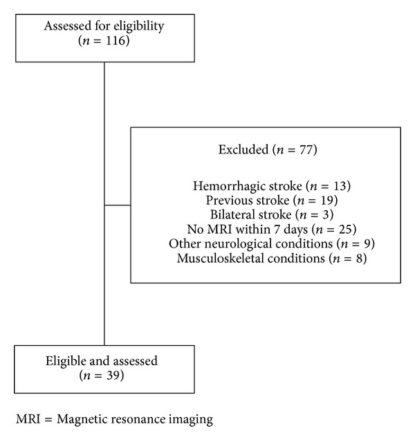
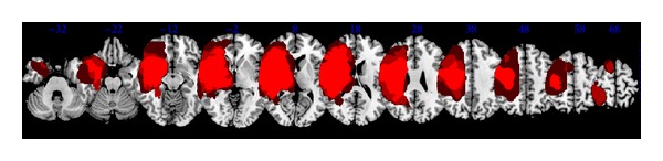
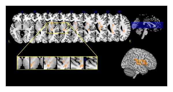
Association between the site of brain injury and poststroke spasticity is poorly understood. The present study investigated whether lesion analysis could document brain regions associated with the development of severe upper limb poststroke spasticity. A retrospective analysis was conducted on 39 chronic stroke patients. Spasticity was assessed at the affected upper limb with the modified Ashworth scale (shoulder, elbow, wrist, and fingers). Brain lesions were traced from magnetic resonance imaging performed within the first 7 days after stroke and region of interest images were generated. The association between severe upper limb spasticity (modified Ashworth scale ≥ 2) and lesion location was determined with the voxel-based lesion-symptom mapping method implemented in MRIcro software. Colored maps representing the z statistics were generated and overlaid onto the automated anatomical labeling and the Johns Hopkins University white matter templates provided with MRIcron. Thalamic nuclei were identified with the Talairach Daemon software. Injuries to the insula, the thalamus, the basal ganglia, and white matter tracts (internal capsule, corona radiata, external capsule, and superior longitudinal fasciculus) were significantly associated with severe upper limb poststroke spasticity. Further advances in our understanding of the neural correlates of spasticity may lead to early targeted rehabilitation when key regions are damaged.
Figures
References
-
- Lance JW. The control of muscle tone, reflexes, and movement: Robert Wartenberg lecture. Neurology. 1980;30(12):1303–1313. - PubMed
-
- Wissel J, Manack A, Brainin M. Toward an epidemiology of poststroke spasticity. Neurology. 2013;80(3, supplement 2):S13–S19. - PubMed
-
- Urban PP, Wolf T, Uebele M, et al. Occurence and clinical predictors of spasticity after ischemic stroke. Stroke. 2010;41(9):2016–2020. - PubMed
-
- Leathley MJ, Gregson JM, Moore AP, Smith TL, Sharma AK, Watkins CL. Predicting spasticity after stroke in those surviving to 12 months. Clinical Rehabilitation. 2004;18(4):438–443. - PubMed
Publication types
MeSH terms
LinkOut - more resources
Full Text Sources
Other Literature Sources
Medical
