Preliminary experience with small animal SPECT imaging on clinical gamma cameras
- PMID: 24963478
- PMCID: PMC4053230
- DOI: 10.1155/2014/369509
Preliminary experience with small animal SPECT imaging on clinical gamma cameras
Abstract
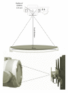
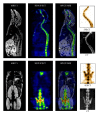
The traditional lack of techniques suitable for in vivo imaging has induced a great interest in molecular imaging for preclinical research. Nevertheless, its use spreads slowly due to the difficulties in justifying the high cost of the current dedicated preclinical scanners. An alternative for lowering the costs is to repurpose old clinical gamma cameras to be used for preclinical imaging. In this paper we assess the performance of a portable device, that is, working coupled to a single-head clinical gamma camera, and we present our preliminary experience in several small animal applications. Our findings, based on phantom experiments and animal studies, provided an image quality, in terms of contrast-noise trade-off, comparable to dedicated preclinical pinhole-based scanners. We feel that our portable device offers an opportunity for recycling the widespread availability of clinical gamma cameras in nuclear medicine departments to be used in small animal SPECT imaging and we hope that it can contribute to spreading the use of preclinical imaging within institutions on tight budgets.
Figures


Similar articles
-
Development of a variable-radius pinhole SPECT system with a portable gamma camera.Rev Esp Med Nucl. 2011 Sep-Oct;30(5):286-91. doi: 10.1016/j.remn.2011.03.004. Epub 2011 Jun 2. Rev Esp Med Nucl. 2011. PMID: 21640439
-
Small animal imaging with multi-pinhole SPECT.Methods. 2009 Jun;48(2):83-91. doi: 10.1016/j.ymeth.2009.03.015. Epub 2009 Mar 26. Methods. 2009. PMID: 19328232 Review.
-
Use of a compact pixellated gamma camera for small animal pinhole SPECT imaging.Ann Nucl Med. 2006 Jul;20(6):409-16. doi: 10.1007/BF03027376. Ann Nucl Med. 2006. PMID: 16922469
-
In vivo quantification of renal function in mice using clinical gamma cameras.Phys Med. 2015 May;31(3):242-7. doi: 10.1016/j.ejmp.2015.01.013. Epub 2015 Feb 26. Phys Med. 2015. PMID: 25726477
-
Ultra-high-resolution imaging of small animals: implications for preclinical and research studies.J Nucl Cardiol. 1999 May-Jun;6(3):332-44. doi: 10.1016/s1071-3581(99)90046-6. J Nucl Cardiol. 1999. PMID: 10385189 Review.
Cited by
-
Preclinical molecular imaging: development of instrumentation for translational research with small laboratory animals.Einstein (Sao Paulo). 2016 Jul-Sep;14(3):408-414. doi: 10.1590/S1679-45082016AO3696. Einstein (Sao Paulo). 2016. PMID: 27759832 Free PMC article.
-
Design and Evaluation of a Portable Pinhole SPECT System for 177Lu Imaging: Monte Carlo Simulations and Experimental Study.Diagnostics (Basel). 2025 May 30;15(11):1387. doi: 10.3390/diagnostics15111387. Diagnostics (Basel). 2025. PMID: 40506959 Free PMC article.
-
Multimodal Molecular Imaging: Current Status and Future Directions.Contrast Media Mol Imaging. 2018 Jun 5;2018:1382183. doi: 10.1155/2018/1382183. eCollection 2018. Contrast Media Mol Imaging. 2018. PMID: 29967571 Free PMC article. Review.
References
-
- Herschman HR. Molecular imaging: looking at problems, seeing solutions. Science. 2003;302(5645):605–608. - PubMed
-
- Willmann JK, van Bruggen N, Dinkelborg LM, Gambhir SS. Molecular imaging in drug development. Nature Reviews Drug Discovery. 2008;7(7):591–607. - PubMed
-
- Beekman F, van der Have F. The pinhole: gateway to ultra-high-resolution three-dimensional radionuclide imaging. European Journal of Nuclear Medicine and Molecular Imaging. 2007;34(2):151–161. - PubMed
-
- MacDonald LR, Patt BE, Iwanczyk JS, et al. Pinhole SPECT of mice using the LumaGEM gamma camera. IEEE Transactions on Nuclear Science. 2001;48(3):830–836.
-
- Funk T, Després P, Barber WC, Shah KS, Hasegawa BH. A multipinhole small animal SPECT system with submillimeter spatial resolution. Medical Physics. 2006;33(5):1259–1268. - PubMed
Publication types
MeSH terms
LinkOut - more resources
Full Text Sources
Other Literature Sources

