

Review
doi: 10.1155/2014/191847.
Epub 2014 May 14.
Spectral-domain optical coherence tomography for macular edema
Affiliations
- PMID: 24963500
- PMCID: PMC4053271
- DOI: 10.1155/2014/191847
Item in Clipboard
Review
Spectral-domain optical coherence tomography for macular edema
ScientificWorldJournal.
2014.
Abstract
Optical coherence tomography (OCT) is a rapid noncontact method that allows in vivo imaging of the retina and it has become an important component in clinical practice. OCT is a useful ancillary tool for assessing retinal diseases because of its ability to provide cross-sectional retinal images and quantitatively analyze retinal morphology. The introduction of spectral-domain OCT provided major improvements in image acquisition speed and image resolution. Future studies will address how these major technologic advances will impact the use of OCT in research and clinical practice.
Figures
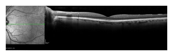
A spectral domain optical coherence tomography line scan of a normal eye.
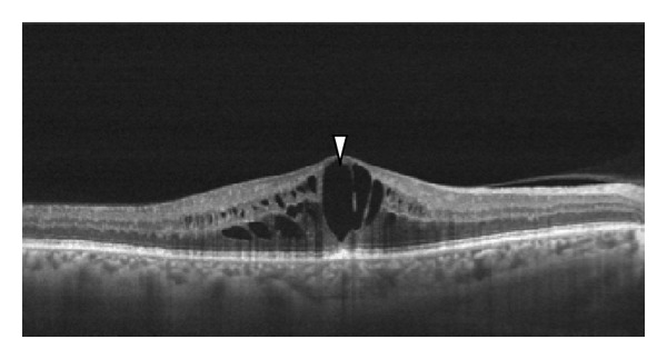
Cystoid macular edema can be seen clearly on OCT scans as multiple circular cystic spaces in the retina, indicating intraretinal edema (white arrowhead). The cystic spaces are round and originate around the outer plexiform layer.

An OCT horizontal line scan of a 62-year-old man with diabetic retinopathy and macular edema-intraretinal cysts (white arrowhead).
An OCT horizontal line scan of a woman with diabetes, with a juxtafoveal accumulation of hard exudates (white arrowhead) and substantial fluid at the level of outer plexiform layer (yellow arrowhead). Diffuse hyperreflective hard exsudates can also be seen (red arrowhead).
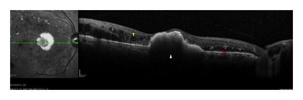
A line scan of an eye with macular edema secondary to an active exudative AMD. The technique allows for visualization of the cystic spaces (white arrowhead) and other changes in the retinal layers. Note the hyperreflective layer underneath the neurosensory retina suggestive of the neovascular membrane complex (yellow arrowhead).
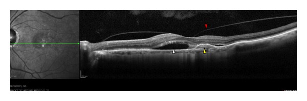
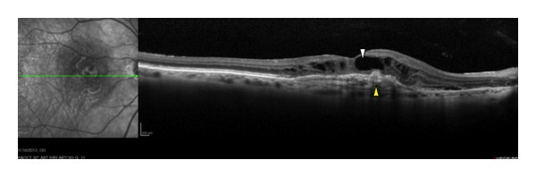
An OCT line scan of a 73-year-old man with exudative AMD. The white arrowhead shows that the hyporeflective space below the neurosensory retina is clearly visible, suggesting the presence of fluid. Yellow arrowhead represents a hemorrhagic detachment of the retinal pigment epithelium (PED) and vitreomacular traction in addition to vitreous alterations (red arrowhead).

Macular edema in a 75-year-old woman with CRVO. There are several cystic spaces in the retinal layers (white arrowhead), although the foveal depression is preserved (yellow arrowhead).
References
-
- Swanson EA, Izatt JA, Hee MR, et al. In vivo retinal imaging by optical coherence tomography. Optics Letters. 1993;18(21):1864–1869. - PubMed
-
- Al-Latayfeh MM, Sun JK, Aiello LP. Ocular coherence tomography and diabetic eye disease. Seminars in Ophthalmology. 2010;25(5-6):192–197. - PubMed
-
- Wolf S, Wolf-Schnurrbusch U. Spectral-domain optical coherence tomography use in macular diseases: a review. Ophthalmologica. 2010;224(6):333–340. - PubMed
-
- Schimel AM, Fisher YL, Flynn HW., Jr. Optical coherence tomography in the diagnosis and management of diabetic macular edema: time-domain versus spectral-domain. Ophthalmic Surgery, Lasers and Imaging. 2011;42:S41–S55. - PubMed
Publication types
MeSH terms
LinkOut - more resources
Full Text Sources
Other Literature Sources
Medical
