

Maternal pravastatin prevents altered fetal brain development in a preeclamptic CD-1 mouse model
- PMID: 24963809
- PMCID: PMC4071009
- DOI: 10.1371/journal.pone.0100873
Maternal pravastatin prevents altered fetal brain development in a preeclamptic CD-1 mouse model
Abstract
Objective: Using an animal model, we have previously shown that preeclampsia results in long-term adverse neuromotor outcomes in the offspring, and this phenotype was prevented by antenatal treatment with pravastatin. This study aims to localize the altered neuromotor programming in this animal model and to evaluate the role of pravastatin in its prevention.
Materials and methods: For the preeclampsia model, pregnant CD-1 mice were randomly allocated to injection of adenovirus carrying sFlt-1 or its control virus carrying mFc into the tail vein. Thereafter they received pravastatin (sFlt-1-pra "experimental group") or water (sFlt-1 "positive control") until weaning. The mFc group ("negative control") received water. Offspring at 6 months of age were sacrificed, and whole brains underwent magnetic resonance imaging (MRI). MRIs were performed using an 11.7 Tesla vertical bore MRI scanner. T2 weighted images were acquired to evaluate the volumes of 28 regions of interest, including areas involved in adaptation and motor, spatial and sensory function. Cytochemistry and cell quantification was performed using neuron-specific Nissl stain. One-way ANOVA with multiple comparison testing was used for statistical analysis.
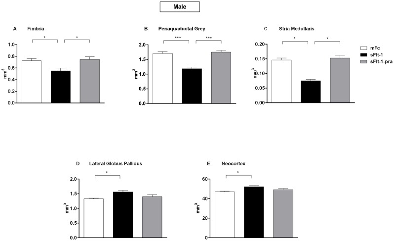
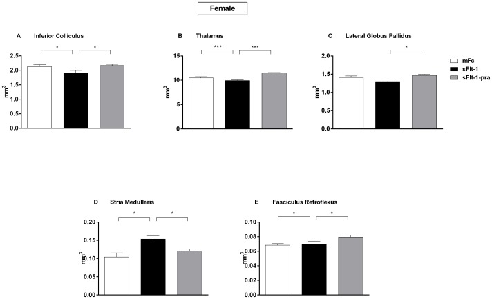
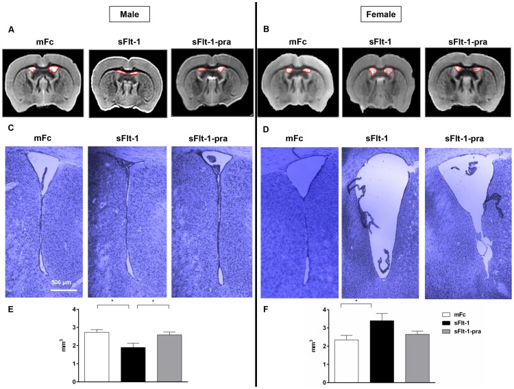
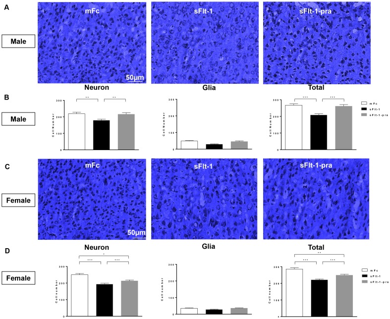
Results: Compared with control offspring, male sFlt-1 offspring have decreased volumes in the fimbria, periaquaductal gray, stria medullaris, and ventricles and increased volumes in the lateral globus pallidus and neocortex; however, female sFlt-1 offspring showed increased volumes in the ventricles, stria medullaris, and fasciculus retroflexus and decreased volumes in the inferior colliculus, thalamus, and lateral globus pallidus. Neuronal quantification via Nissl staining exhibited decreased cell counts in sFlt-1 offspring neocortex, more pronounced in males. Prenatal pravastatin treatment prevented these changes.
Conclusion: Preeclampsia alters brain development in sex-specific patterns, and prenatal pravastatin therapy prevents altered neuroanatomic programming in this animal model.
Conflict of interest statement
Figures
Similar articles
-
The effect of prenatal pravastatin treatment on altered fetal programming of postnatal growth and metabolic function in a preeclampsia-like murine model.Am J Obstet Gynecol. 2014 Jun;210(6):542.e1-7. doi: 10.1016/j.ajog.2014.01.010. Epub 2014 Jan 8. Am J Obstet Gynecol. 2014. PMID: 24412113
-
Effects of pravastatin on angiogenic and placental hypoxic imbalance in a mouse model of preeclampsia.Reprod Sci. 2014 Jan;21(1):138-45. doi: 10.1177/1933719113492207. Epub 2013 Jun 7. Reprod Sci. 2014. PMID: 23749761
-
Pravastatin Effects on Placental Prosurvival Molecular Pathways in a Mouse Model of Preeclampsia.Reprod Sci. 2016 Nov;23(11):1593-1599. doi: 10.1177/1933719116648218. Epub 2016 May 11. Reprod Sci. 2016. PMID: 27170663
-
Therapeutic potential of statins and the induction of heme oxygenase-1 in preeclampsia.J Reprod Immunol. 2014 Mar;101-102(100):153-160. doi: 10.1016/j.jri.2013.12.120. Epub 2014 Jan 16. J Reprod Immunol. 2014. PMID: 24503248 Free PMC article. Review.
-
Pravastatin to treat and prevent preeclampsia. Preclinical and clinical studies.J Reprod Immunol. 2017 Nov;124:15-20. doi: 10.1016/j.jri.2017.09.009. Epub 2017 Sep 29. J Reprod Immunol. 2017. PMID: 29028516 Review.
Cited by
-
Systemic Maternal Human sFLT1 Overexpression Leads to an Impaired Foetal Brain Development of Growth-Restricted Foetuses upon Experimental Preeclampsia.Oxid Med Cell Longev. 2022 Jun 2;2022:3024032. doi: 10.1155/2022/3024032. eCollection 2022. Oxid Med Cell Longev. 2022. PMID: 35693702 Free PMC article.
-
The Role of Statins in Prevention of Preeclampsia: A Promise for the Future?Front Pharmacol. 2017 May 5;8:247. doi: 10.3389/fphar.2017.00247. eCollection 2017. Front Pharmacol. 2017. PMID: 28529486 Free PMC article. Review.
-
Pravastatin to prevent recurrent fetal death in massive perivillous fibrin deposition of the placenta (MPFD).J Matern Fetal Neonatal Med. 2016 Mar;29(6):855-62. doi: 10.3109/14767058.2015.1022864. Epub 2015 Apr 20. J Matern Fetal Neonatal Med. 2016. PMID: 25893545 Free PMC article.
-
Adult Pgf-/- mice behaviour and neuroanatomy are altered by neonatal treatment with recombinant placental growth factor.Sci Rep. 2019 Jun 26;9(1):9285. doi: 10.1038/s41598-019-45824-6. Sci Rep. 2019. PMID: 31243296 Free PMC article.
-
The Impact of Increased Maternal sFlt-1/PlGF Ratio on Motor Outcome of Preterm Infants.Front Endocrinol (Lausanne). 2022 Jun 30;13:913514. doi: 10.3389/fendo.2022.913514. eCollection 2022. Front Endocrinol (Lausanne). 2022. PMID: 35846340 Free PMC article.
References
-
- American College of Obstetricians and Gynecologists (2002) Diagnosis and management of preeclampsia and eclampsia. ACOG Practice Bulletin No. 33. Obstet Gynecol 99: 159–167. - PubMed
-
- Redman CW, Sargent IL (2005) Latest advances in understanding preeclampsia. Science 308: 1592–1594. - PubMed
-
- Levine RJ, Maynard SE, Qian C, Lim KH, England LJ, et al. (2004) Circulating angiogenic factors and the risk of preeclampsia. N Engl J Med 350: 672–683. - PubMed
-
- Lu F, Bytautiene E, Tamayo E, Gamble P, Anderson GD, et al. (2007) Gender-specific effect of over-expression of sFlt-1 in pregnant mice on fetal programming of blood pressure in the offspring later in life. Am J Obstet Gynecol 197: 418e1–418e5. - PubMed
Publication types
MeSH terms
Substances
Grants and funding
LinkOut - more resources
Full Text Sources
Other Literature Sources
Medical
Research Materials
