. 2015 May;41(5):1475-85.
doi: 10.1002/jmri.24681. Epub 2014 Jun 25.

Measurements of wall shear stress and aortic pulse wave velocity in swine with familial hypercholesterolemia

Affiliations

Measurements of wall shear stress and aortic pulse wave velocity in swine with familial hypercholesterolemia

Andrew L Wentland et al. J Magn Reson Imaging. 2015 May.

Abstract

Purpose: To assess measurements of pulse wave velocity (PWV) and wall shear stress (WSS) in a swine model of atherosclerosis.

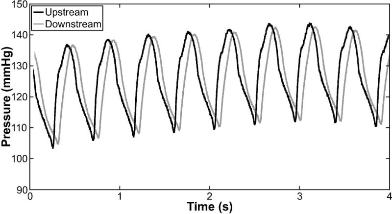
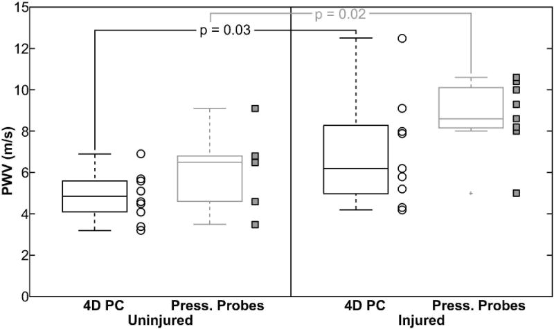
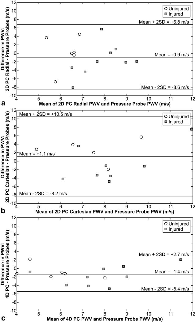
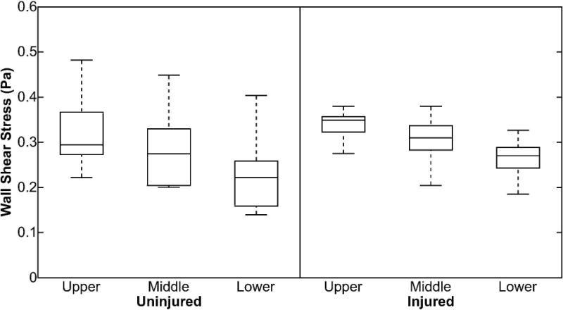
Materials and methods: Nine familial hypercholesterolemic (FH) swine with angioplasty balloon catheter-induced atherosclerotic lesions to the abdominal aorta (injured group) and 10 uninjured FH swine were evaluated with a 4D phase contrast (PC) magnetic resonance imaging (MRI) acquisition, as well as with radial and Cartesian 2D PC acquisitions, on a 3T MR scanner. PWV values were computed from the 2D and 4D PC techniques, compared between the injured and uninjured swine, and validated against reference standard pressure probe-based PWV measurements. WSS values were also computed from the 4D PC MRI technique and compared between injured and uninjured groups.

Results: PWV values were significantly greater in the injured than in the uninjured groups with the 4D PC MRI technique (P = 0.03) and pressure probes (P = 0.02). No significant differences were found in PWV between groups using the 2D PC techniques (P = 0.75-0.83). No significant differences were found for WSS values between the injured and uninjured groups.

Conclusion: The 4D PC MRI technique provides a promising means of evaluating PWV and WSS in a swine model of atherosclerosis, providing a potential platform for developing the technique for the early detection of atherosclerosis.

Keywords: 4D PC MRI; PWV; atherosclerosis; familial hypercholesterolemia; pulse wave velocity; wall shear stress.


Figures

Figure 1
Figure 1
Angiograms provided by C-arm computed tomography. Balloon catheters were deployed within one group of pigs and used to denude the endothelium in two locations, as identified by the yellow boxes in the suprarenal and infrarenal abdominal aorta (a). Part b demonstrates the prescription of the five 2D planes that were used for the acquisition of radial and Cartesian 2D phase contrast (PC) data sets. Part b also depicts the field of view prescribed for the acquisition of the 4D PC data set. For wall shear stress analysis, values were averaged over three sections (c)—the upper (suprarenal aorta), middle (pararenal aorta), and lower (infrarenal aorta) sections. In some angiograms, irregularities could be identified in the areas of injury in the abdominal aorta (d).
Figure 2
Figure 2
Pressure waveforms provided by two fiber optic pressure sensors that sampled at a frequency of 1 kHz. The probes were separated by 10 cm and placed in the abdominal aorta of swine. The upstream probe was approximately 5 cm superior to the renal arteries, whereas the downstream probe was approximately 5 cm inferior to the renal arteries. Drift in the signal was due to respiration.
Figure 3
Figure 3
Scatter and box plots of pulse wave velocity (PWV) values measured from a 4D phase contrast (4D PC) technique, as well as gold standard fiber optic intravascular pressure probes, in swine with and without injury to the abdominal aorta. Both boxes and individual points are shown for each group. PWV values in the injured group were significantly greater than values in the uninjured group for both the 4D PC technique (p = 0.03) and the pressure probe measurements (p = 0.02). Press. = Pressure.
Figure 4
Figure 4
Bland-Altman analysis comparing pulse wave velocity (PWV) measurements derived from radial 2D PC (a), Cartesian 2D PC (b), and 4D PC (c) to gold standard pressure probe-based measurements of PWV. The mean bias (±2 SD) was −0.9 ± 7.7, +1.1 ± 9.4, and −1.4 ± 4.1 m/s for the radial 2D PC, Cartesian 2D PC, and 4D PC techniques, respectively.
Figure 5
Figure 5
Box plots of wall shear stress in uninjured and injured swine, in which the upper segment corresponds to the suprarenal abdominal aorta, the middle segment corresponds to the pararenal aorta, and the lower segment corresponds to the infrarenal abdominal aorta. In the injured group, the upper and lower segments align with areas of injury with a balloon catheter.
Figure 6
Figure 6
Hematoxylin and eosin (H&E) stain of slices extracted from the infrarenal abdominal aorta of uninjured (a,b) and injured (c,d) FH swine with a balloon catheter-induced injury. Each of the four slices shown was extracted from a different animal. Most swine with an injured aorta demonstrated substantial plaque development and intimal wall thickening (c,d). Uninjured swine demonstrated minimal (b) to no (a) plaque development in the same regions of the aorta.

Similar articles

Cited by

References

    1. Gaziano JM, Wilson PW. Cardiovascular risk assessment in the 21st century. JAMA. 2012;308(8):816–817. - PubMed
    1. Den Ruijter HM, Peters SA, Anderson TJ, et al. Common carotid intima-media thickness measurements in cardiovascular risk prediction: a meta-analysis. JAMA. 2012;308(8):796–803. - PubMed
    1. Yeboah J, McClelland RL, Polonsky TS, et al. Comparison of novel risk markers for improvement in cardiovascular risk assessment in intermediate-risk individuals. JAMA. 2012;308(8):788–795. - PMC - PubMed
    1. Schmermund A, Erbel R. Unstable coronary plaque and its relation to coronary calcium. Circulation. 2001;104(14):1682–1687. - PubMed
    1. Taylor AJ, Burke AP, O’Malley PG, et al. A comparison of the Framingham risk index, coronary artery calcification, and culprit plaque morphology in sudden cardiac death. Circulation. 2000;101(11):1243–1248. - PubMed

Publication types