. 2014 Jun 25;9(6):e100002.
doi: 10.1371/journal.pone.0100002. eCollection 2014.

Death and transfiguration in static Staphylococcus epidermidis cultures

Affiliations

Death and transfiguration in static Staphylococcus epidermidis cultures

Christoph Schaudinn et al. PLoS One. .

Abstract

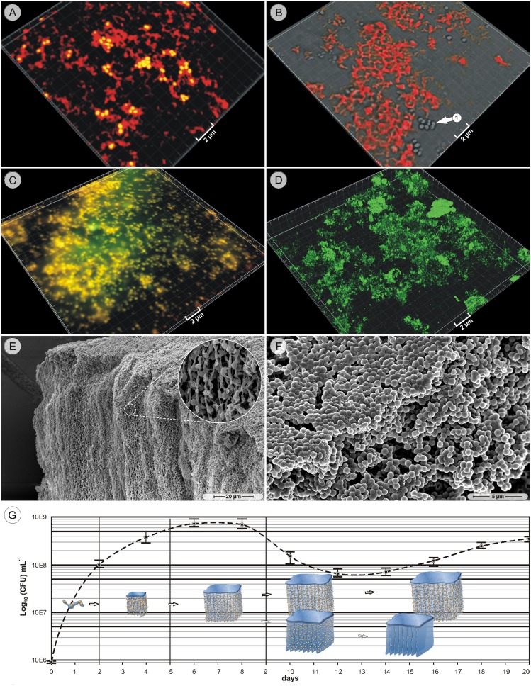
The overwhelming majority of bacteria live in slime embedded microbial communities termed biofilms, which are typically adherent to a surface. However, when several Staphylococcus epidermidis strains were cultivated in static liquid cultures, macroscopic aggregates were seen floating within the broth and also sedimented at the test tube bottom. Light- and electron microscopy revealed that early-stage aggregates consisted of bacteria and extracellular matrix, organized in sheet-like structures. Perpendicular under the sheets hung a network of periodically arranged, bacteria-associated strands. During the extended cultivation, the strands of a subpopulation of aggregates developed into cross-connected wall-like structures, in which aligned bacteria formed the walls. The resulting architecture had a compartmentalized appearance. In late-stage cultures, the wall-associated bacteria disintegrated so that, henceforth, the walls were made of the coalescing remnants of lysed bacteria, while the compartment-like organization remained intact. At the same time, the majority of strand-containing aggregates with associated culturable bacteria continued to exist. These observations indicate that some strains of Staphylococcus epidermidis are able to build highly sophisticated structures, in which a subpopulation undergoes cell lysis, presumably to provide continued access to nutrients in a nutrient-limited environment, whilst maintaining structural integrity.


Conflict of interest statement

Competing Interests: D. H. Robinson holds the US Patent No; 8,187,857 “Multicellular organisms derived from normal/nondiseased mammalian tissues”. The authors hereby declare that the affiliation of D. H. Robinson with deNovo Biologic LLC does not alter in any way the authors' adherence to PLOS ONE policies on sharing data and materials. The authors furthermore declare that D. H. Robinson’s holding of the US Patent No; 8,187,857 “Multicellular organisms derived from normal/nondiseased mammalian tissues” does not alter in any way the authors’ adherence to PLOS ONE policies on sharing data and materials.

Figures

Figure 1
Figure 1. Early stage structures.
A–C. Camera pictures, MH strain: floating and sedimented macroscopic aggregates of a 1-day culture. D. Scanning electron microscopy (SEM) picture, MH strain, 1-day culture: bacteria (arrow 1) and matrix elements (arrow 2) form small aggregates. E. SEM picture, 3-day culture, MH strain: a fibrous sheet (region 1) and short strands (arrow 2) form larger aggregates. F. SEM picture, 5-day culture, MH strain: aggregates consist of solid sheets (region 1) and subjacent a network of strands (region 2). G. SEM picture, 5-day culture, MH strain: predominantly parallel orientation of the strand network. H. SEM picture, 5-day culture: bacteria-associated strands (arrow 1), cross-connected by fibers (arrow 2). I. Confocal laser scanning microscopy (cLSM) picture, MH strain, 10-day culture: strands and associated particulate material (arrows 1, 2) are stained with Concanavalin A; bacteria are labeled with Syto59 (arrow 3). J. SEM picture, 10-day culture, MH strain: almost solid, parallel strands with cross-connecting fibers.
Figure 2
Figure 2. Compartmentalization.
A. SEM picture, 10-day culture, MH strain: solid, parallel, wall-like structures. B. SEM picture, 14-day culture, MH strain: the top sheet (region 1) is situated on vertically stacked walls (region 2). C. cLSM transmission mode, 14-day culture, MH strain: a dense, horizontal top plate (region 1) is located on top of vertically stacked walls (region 2). D. SEM picture, 14-day culture, MH strain: compartmentalized structure of aligned walls and cross-walls without top sheet. E. cLSM transmission mode, 14-day culture, MH strain: aligned bacteria form compartment walls. F. SEM picture, 14-day culture, MH strain: matrix (arrow 1) embedded bacteria (arrow 2) form compartment walls. G. cLSM picture, 14-day culture, MH strain: LIVE/DEAD staining of compartment wall forming bacteria show varying stages of membrane integrity (arrows). H. SEM picture, 14-day culture, MH strain: compartment structure with abundant bacteria (region 1) adjacent to an area with bacteria-depleted compartments.
Figure 3
Figure 3. Late stage structures.
A. SEM picture, 14-day culture, MH strain: the thin compartment walls contain only few bacteria. B. Transmission electron microscopy (TEM) picture, 14-day culture, MH strain: the few bacteria (arrow 1) are a part of otherwise in ‘empty’ walls (arrow 2). C. TEM picture, 14-day culture, MH strain: an intact bacterium (arrow 1) and a partly disintegrated bacterium (arrow 2) are both integrated in the wall structure (arrow 3). D. TEM picture, 14-day culture, MH strain: the bare walls consist of fine, dispersed fibers (arrow 1) and intensely stained dots (arrow 2). E. SEM picture, 14-day culture, MH strain: empty compartment walls. F. SEM picture, 14-day culture, MH strain: bacteria depleted compartment walls. G. SEM picture: 14-day old cultures (MH strain) showed large compartmentalized areas. H. cLSM transmission mode, 14-day culture (MH strain): ‘empty’ compartment walls under hydrated conditions.
Figure 4
Figure 4. Specific staining, time-course and development model.
A. cLSM picture, 14-day culture, strain #49134: the EUB338 probe labels the compartment structure (red), while the bacteria are double-stained by the EUB338 probe and Syto59 (yellow). B. cLSM picture, 14-day culture, MH strain: Concanavalin A stains the compartment structure, but not the bacteria (arrow 1). C. cLSM picture, 14-day culture, strain #49134: the Sypro protein stain (green) and the Syto59 nucleic acid stain label both the bacteria (yellow) but not the compartment structure. D. cLSM picture, 14-day culture, strain #49134: the lipophilic stain Nile Red (green) does not label compartment walls, only diffuse biofilm matrix. E. SEM picture, 28-day culture, MH strain: huge aggregates with intact bacteria and strands continued to be present in long-time cultures. F. SEM picture, MH strain, overnight grown colony on agar. G. The model depicts the main development stages and their timely appearances in the context with the CFU time-course (MH strain).

References

    1. Costerton JW, Lewandowski Z, Caldwell DE, Korber DR, Lappin-Scott HM (1995) Microbial biofilms. Annu Rev Microbiol 49: 711–745. - PubMed
    1. Wimpenny J, Manz W, Szewzyk U (2000) Heterogeneity in biofilms. FEMS Microbiol Rev 24: 661–671. - PubMed
    1. Bockelmann U, Manz W, Neu TR, Szewzyk U (2002) Investigation of lotic microbial aggregates by a combined technique of fluorescent in situ hybridization and lectin-binding-analysis. J Microbiol Methods 49: 75–87. - PubMed
    1. Monds RD, O’Toole GA (2009) The developmental model of microbial biofilms: ten years of a paradigm up for review. Trends Microbiol 17: 73–87. - PubMed
    1. Allesen-Holm M, Barken KB, Yang L, Klausen M, Webb JS, et al. (2006) A characterization of DNA release in Pseudomonas aeruginosa cultures and biofilms. Mol Microbiol 59: 1114–1128. - PubMed

Publication types

MeSH terms

LinkOut - more resources