. 2014 Aug;9(8):1013-22.
doi: 10.1002/biot.201300483.

Construction of pH-sensitive Her2-binding IgG1-Fc by directed evolution

Affiliations
Free PMC article

Construction of pH-sensitive Her2-binding IgG1-Fc by directed evolution

Michael W Traxlmayr et al. Biotechnol J. 2014 Aug.
Free PMC article

Abstract

For most therapeutic proteins, a long serum half-life is desired. Studies have shown that decreased antigen binding at acidic pH can increase serum half-life. In this study, we aimed to investigate whether pH-dependent binding sites can be introduced into antigen binding crystallizable fragments of immunoglobulin G1 (Fcab). The C-terminal structural loops of an Fcab were engineered for reduced binding to the extracellular domain of human epidermal growth factor receptor 2 (Her2-ECD) at pH 6 compared to pH 7.4. A yeast-displayed Fcab-library was alternately selected for binding at pH 7.4 and non-binding at pH 6.0. Selected Fcab variants showed clear pH-dependent binding to soluble Her2-ECD (decrease in affinity at pH 6.0 compared to pH 7.4) when displayed on yeast. Additionally, some solubly expressed variants exhibited pH-dependent interactions with Her2-positive cells whereas their conformational and thermal stability was pH-independent. Interestingly, two of the three Fcabs did not contain a single histidine mutation but all of them contained variations next to histidines that already occurred in loops of the lead Fcab. The study demonstrates that yeast surface display is a valuable tool for directed evolution of pH-dependent binding sites in proteins.

Keywords: Antibody engineering; Directed evolution; Fcab; Yeast surface display; pH-depending binding.


Figures

Figure 1
Figure 1
Front and side view of the structure of human IgG1-Fc (PDB-ID 1OQO). The two chains of the homodimeric protein are depicted in light and dark gray, respectively. Each chain comprises a CH2 (top) and a CH3 domain (bottom). The randomized regions in the C-terminal structural loops of the CH3 domains are depicted in red (AB-loop) and blue (EF-loop). For sequence information, see Table 1.
Figure 2
Figure 2
(A) Selection strategy for the generation of pH-dependent binders. A yeast displayed Fcab library was incubated with 3 nM biotinylated Her2-ECD, followed by labeling with SA-PE and anti-CH2-FITC. The incubations were either performed at pH 7.4 (selection rounds 1, 2, and 5) or at pH 6.0 (rounds 3, 4, and 6). If the staining procedure was done at pH 7.4, the cells were subsequently sorted for binding to Her2, whereas incubation at pH 6.0 was followed by selection of non-binders. Exemplarily, the dot plots from selection rounds 4 (top) and 5 (bottom) are shown. The boxes within the plots indicate the gates that were used for sorting. (B) Analysis of the pH-dependence of Her2-binding of selected Fcab mutants. After six rounds of flow cytometric sorting, individual Fcab-clones were analyzed. Fcabs were displayed on yeast and incubated with 3 nM biotinylated Her2, followed by detection of antigen binding with SA-PE and of the surface display level with anti-Xpress-APC (recognizing an N-terminal expression tag located between Aga2p and the Fcab). Incubation steps were either performed at pH 6.0 (gray lines) or 7.4 (black lines). Only Xpress-positive (i.e. displaying) cells were analyzed. In total, 10 Fcabs that have been enriched during selection were tested for pH-dependent Her2 binding. Only the three best performing clones (P1, P2, and P3) are shown.
Figure 3
Figure 3
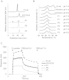
(A) Size exclusion chromatography (SEC) analysis. Ten micrograms of Fc protein [dialyzed in PBS, pH 7.4 (black) or pH 6.0 (gray)] were loaded onto a Superdex 200 column (10 mm × 300 mm, GE Healthcare) at 0.75 mL/min in PBS containing 200 mM NaCl. (B) Differential scanning calorimetry (DSC) analysis. Fcabs were set to a concentration of 5 μM in PBS at pH 6.0 or 7.4. Thermal unfolding was analyzed at a heating rate of 1°C min–1. (C) Binding of Fcabs to FcRn. FcRn was immobilized onto an SPR chip. The coated chip was floated with 10 μg/mL Fcab (in PBS pH 6.0). Finally, pH-dependent dissociation was monitored in PBS pH 6.0, followed by PBS pH 7.4.
Figure 4
Figure 4
pH-dependent binding of Fcabs to Her2 positive cells. The Her2 positive cell line SKBR-3 was incubated with various Fcab concentrations. Subsequently, surface bound Fcab was detected with polyclonal R-phycoerythrin-labeled F(ab')2 fragment directed against human IgG. The incubations were performed at either pH 6.0 (empty) or 7.4 (filled symbols). Mean values ± standard deviations from three independent experiments are shown.

Similar articles

Cited by

References

    1. Kontermann RE. Strategies for extended serum half-life of protein therapeutics. Curr. Opin. Biotechnol. 2011;22:868–876. - PubMed
    1. Maxfield FR, McGraw TE. Endocytic recycling. Nat. Rev. Mol. Cell. Biol. 2004;5:121–132. - PubMed
    1. Igawa T, Ishii S, Tachibana T, Maeda A. Antibody recycling by engineered pH-dependent antigen binding improves the duration of antigen neutralization. Nat. Biotechnol. 2010;28:1203–1207. - PubMed
    1. Chaparro-Riggers J, Liang H, DeVay RM, Bai L. Increasing serum half-life and extending cholesterol lowering in vivo by ngineering antibody with pH-sensitive binding to PCSK9. J. Biol. Chem. 2012;287:11090–11097. - PMC - PubMed
    1. Sarkar CA, Lowenhaupt K, Horan T, Boone TC. Rational cytokine design for increased lifetime and enhanced potency using pH-activated histidine-switching. Nat. Biotechnol. 2002;20:908–913. - PubMed

Publication types

MeSH terms