

Pleomorphic adenoma of lacrimal gland
- PMID: 24964328
- PMCID: PMC3853974
- DOI: 10.1093/jscr/rjt089
Pleomorphic adenoma of lacrimal gland
Abstract
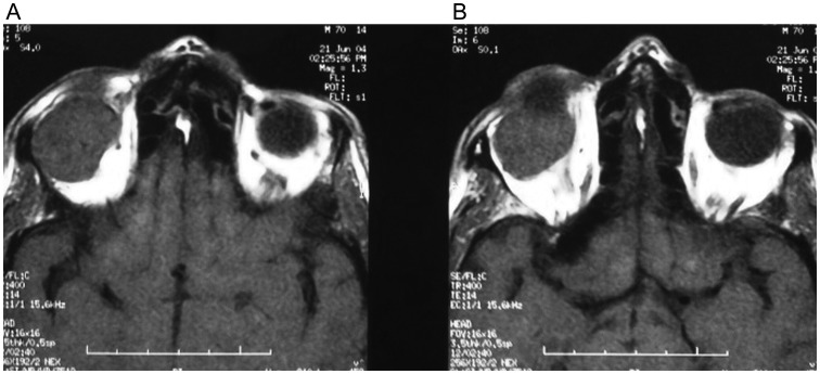
We present a case of a 62-year-old male patient with pleomorphic adenoma and painless solid mass in his right eye. Computerized tomography demonstrated a mass with a diameter of 2.5 cm located in the right lacrimal gland. The mass was removed completely by combined orbitofrontal craniotomy through a transcranial approach. Histopathologic examination revealed pleomorphic adenoma of the lacrimal gland. Orbital tumors originate primarily from vascular, muscle, cartilage, neural tissues, lacrimal glands and lymphoid structures. Five percent of all intraorbital masses originate from the lacrimal gland. Pleomorphic adenoma presents as a painless and slowly growing mass and also as exophthalmoses. Pleomorphic adenoma has a high morbidity. Morbidity increases due to the total displacement of the tumor without its capsule and incisional biopsy for the purpose of diagnosis. The success of the treatment depends on the removal of the tumor with its capsule.
Published by Oxford University Press and JSCR Publishing Ltd. All rights reserved. © The Author 2013.
Figures


References
-
- Font RL, Smith SL, Bryan RG. Malignant epithelial tumors of the lacrimal gland: a clinicopathologic study of 21 cases. Arch Ophthalmol. 1998;116:613–16. - PubMed
-
- Ferry AP, Font RL. Carcinoma metastatic to the orbit. Mod Probl Ophthalmol. 1975;14:377–81. - PubMed
-
- Billroth T. Beobachtungen fiber Geschwulste der Speicheldriisen. Virchow Arch Path Anat. 1859;17:357–75.
-
- Koike T, Sano S, Tajima S, Sakaguchi M, Toyoda N, Yamamoto Y, et al. A case of pleomorphic adenoma of the lacrimal gland. Nippon Keiseigeka Gakkai Kaishi. 1989;9:150–5.
-
- Perzin KH, Jakobiec FA, Livolsi VA, Desjardins L. Lacrimal gland malignant mixed tumors (carcinomas arising in benign mixed tumors) Cancer. 1980;45:2593–606. - PubMed
Publication types
LinkOut - more resources
Full Text Sources
Other Literature Sources
