

Pediatric parafalcine empyemas
- PMID: 24964473
- PMCID: PMC3813702
- DOI: 10.1093/jscr/rjt067
Pediatric parafalcine empyemas
Abstract
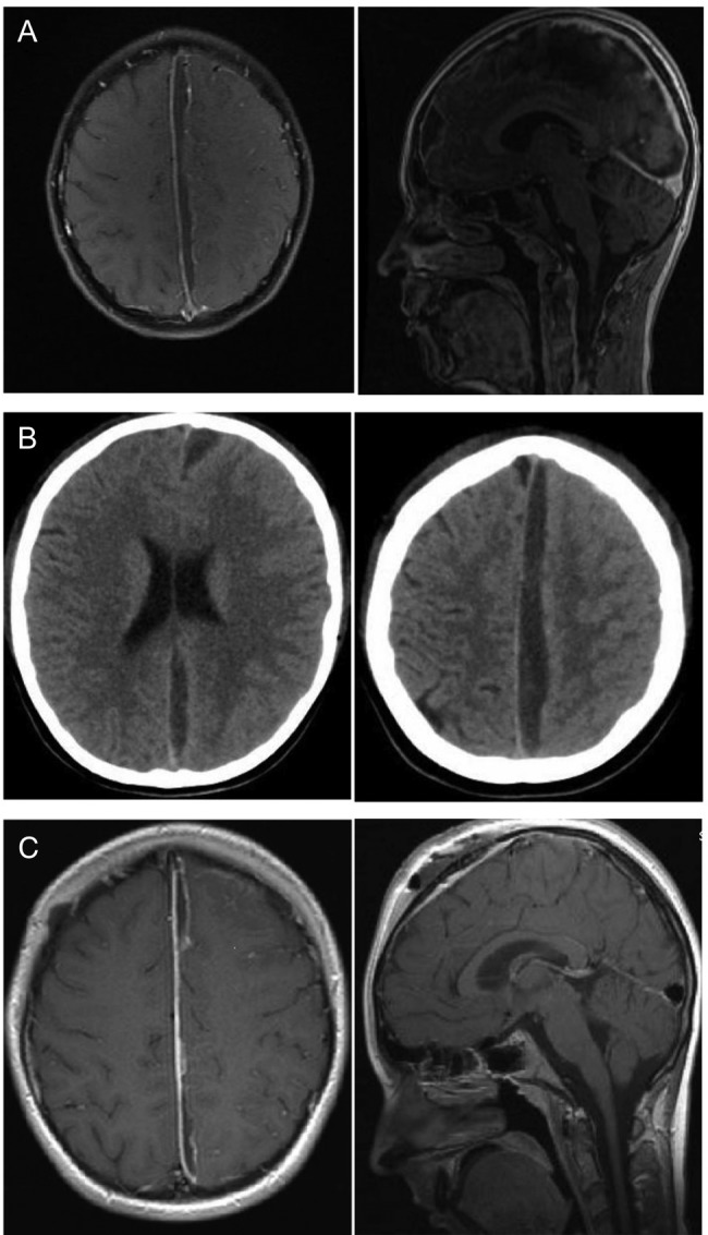
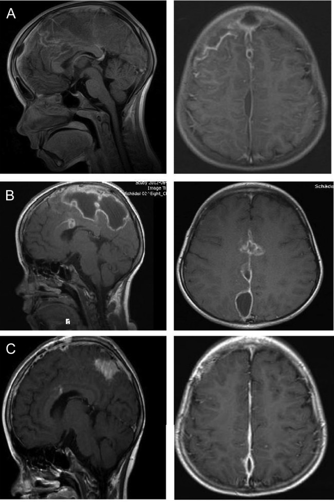
Subdural intracranial empyemas and brain abscesses are a rare complication of bacterial sinusitis. Pediatric parafalcine abscesses are a rare entity with different treatment compared with other brain abscesses. We present two pediatric cases with falcine abscess as a sinusitis complication and introduce our department's treatment management. In addition a review of literature is performed. Surgical cases of our department and their management are compared with the current literature. In our cases, both of the children showed a recurrent empyema after the first surgical treatment and antibiotic therapy. A second surgical evacuation was necessary. The antibiotic therapy was given for 3 months. Short-time follow-up imaging is necessary irrespective of infection parameters in blood and patient's clinical condition. Especially in parafalcine abscesses a second look may be an option and surgical treatment with evacuation of pus is the treatment of choice if abscess remnants are visualized.
Published by Oxford University Press and JSCR Publishing Ltd. All rights reserved. © The Author 2013.
Figures
References
-
- Banerjee AD, Pandey P, Devi BI, Sampath S, Chandramouli BA, et al. Pediatric supratentorial subdural empyemas: a retrospective analysis of 65 cases. Pediatr Neurosurg. 2009;45:11–8. doi:10.1159/000202619. - DOI - PubMed
-
- Salunke PS, Malik V, Kovai P, Mukherjee KK. Falcotentorial subdural empyema: analysis of 10 cases. Acta Neurochir (Wien) 2011;153:164–9. discussion 170 doi:10.1007/s00701-010-0695-5. - DOI - PubMed
-
- Ak HE, Ozkan U, Devecioglu C, Kemaloglu MS. Treatment of subdural empyema by burr hole. Isr J Med Sci. 1996;32:542–4. - PubMed
-
- Dill SR, Cobbs CG, McDonald CK. Subdural empyema: analysis of 32 cases and review. Clin Infect Dis. 1995;20:372–86. doi:10.1093/clinids/20.2.372. - DOI - PubMed
Publication types
LinkOut - more resources
Full Text Sources
Other Literature Sources
