

Involvement of ERCC1 in the pathogenesis of osteoarthritis through the modulation of apoptosis and cellular senescence
- PMID: 24964749
- PMCID: PMC4134687
- DOI: 10.1002/jor.22656
Involvement of ERCC1 in the pathogenesis of osteoarthritis through the modulation of apoptosis and cellular senescence
Abstract
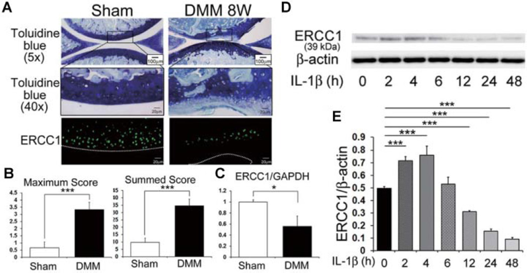
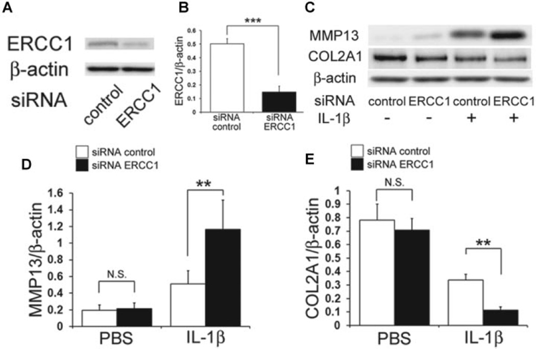
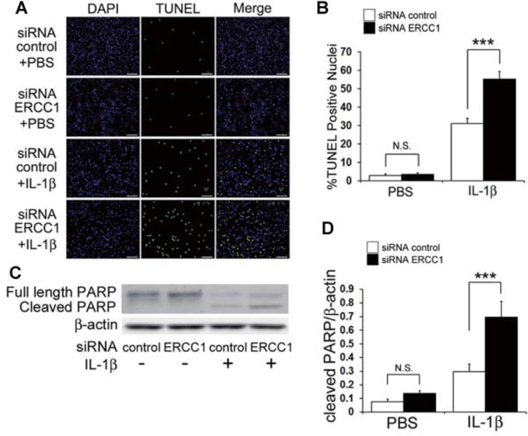
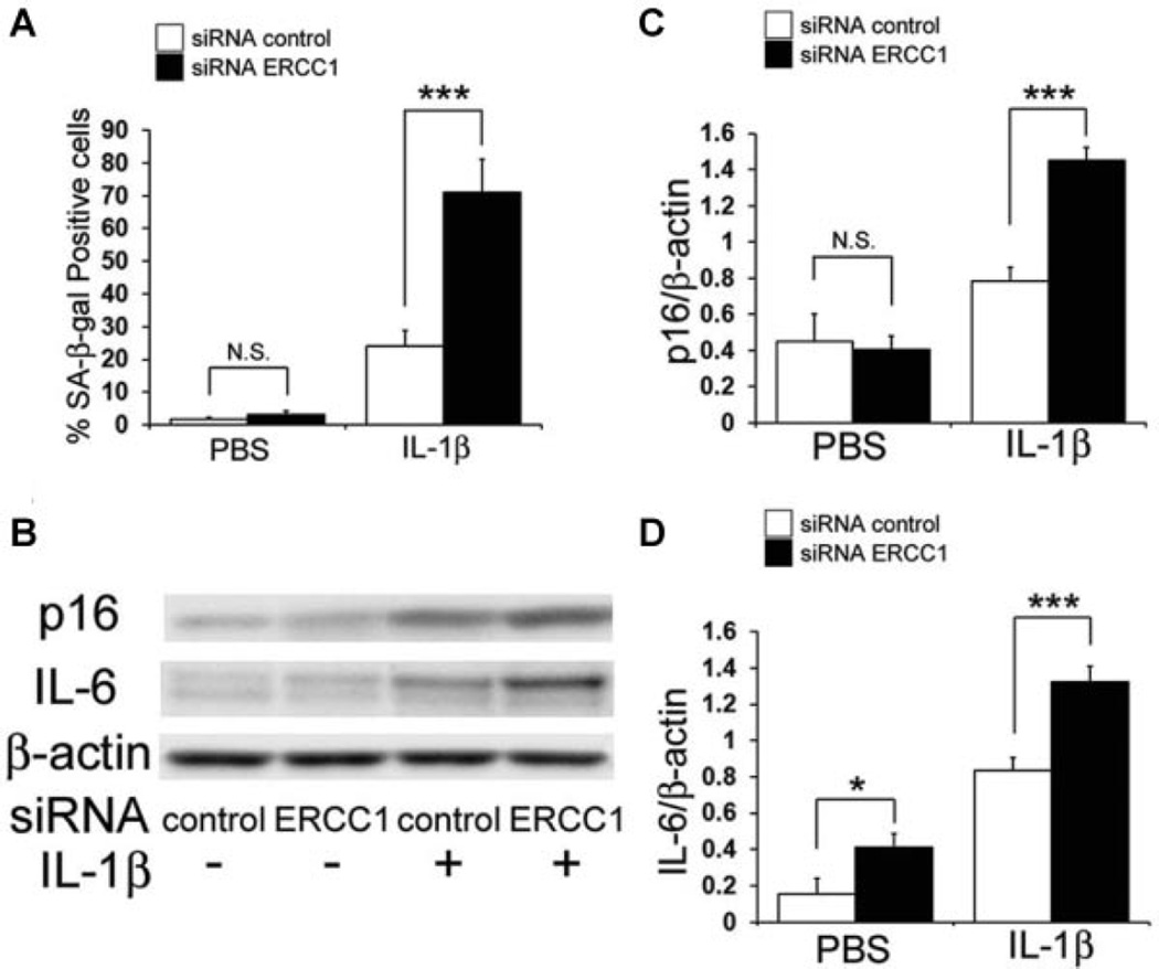
DNA damage is a cause of age related pathologies, including osteoarthritis (OA). Excision repair cross complementation group 1 (ERCC1) is an endonuclease required for DNA damage repair. In this study we investigated the function of ERCC1 in chondrocytes and its association with the pathophysiology of OA. ERCC1 expression in normal and osteoarthritic cartilage was assessed, as were changes in ERCC1 expression in chondrocytes under catabolic stress. Inhibiting ERCC1 in chondrocytes under interleukin-1β stimulation using small interfering RNA (siRNA) was also evaluated. Finally, cellular senescence and apoptosis were examined in relation to ERCC1 function. ERCC1 expression was decreased in OA cartilage and increased within 4 h of exposure to interleukin (IL)-1β, but decreased after 12 h. The inhibition of ERCC1 by siRNA increased the expression of matrix metallopeptidase 13 and decreased collagen type II. ERCC1 inhibition also increased the number of apoptotic and senescent cells. The inhibition of ERCC1 in chondrocytes increased their expression of OA related proteins, apoptosis, cellular senescence, and hypertrophic-like changes which suggest that ERCC1 is critical for protecting human chondrocytes (HCs) from catabolic stresses and provides insights into the pathophysiology of OA and a potential target for its treatment. (191)
Keywords: ERCC1; apoptosis; interleukin (IL)-1β; osteoarthritis; senescence.
© 2014 Orthopaedic Research Society. Published by Wiley Periodicals, Inc.
Conflict of interest statement
Conflict of interest: None.
Figures
Similar articles
-
Deficiency in the DNA repair protein ERCC1 triggers a link between senescence and apoptosis in human fibroblasts and mouse skin.Aging Cell. 2020 Mar;19(3):e13072. doi: 10.1111/acel.13072. Epub 2019 Nov 18. Aging Cell. 2020. PMID: 31737985 Free PMC article.
-
Catabolic stress induces features of chondrocyte senescence through overexpression of caveolin 1: possible involvement of caveolin 1-induced down-regulation of articular chondrocytes in the pathogenesis of osteoarthritis.Arthritis Rheum. 2006 Mar;54(3):818-31. doi: 10.1002/art.21639. Arthritis Rheum. 2006. PMID: 16508959
-
Targeting of chondrocyte plasticity via connexin43 modulation attenuates cellular senescence and fosters a pro-regenerative environment in osteoarthritis.Cell Death Dis. 2018 Dec 5;9(12):1166. doi: 10.1038/s41419-018-1225-2. Cell Death Dis. 2018. PMID: 30518918 Free PMC article.
-
Aging, articular cartilage chondrocyte senescence and osteoarthritis.Biogerontology. 2002;3(5):257-64. doi: 10.1023/a:1020185404126. Biogerontology. 2002. PMID: 12237562 Review.
-
Roles of articular cartilage aging and chondrocyte senescence in the pathogenesis of osteoarthritis.Iowa Orthop J. 2001;21:1-7. Iowa Orthop J. 2001. PMID: 11813939 Free PMC article. Review.
Cited by
-
Ageing and the pathogenesis of osteoarthritis.Nat Rev Rheumatol. 2016 Jul;12(7):412-20. doi: 10.1038/nrrheum.2016.65. Epub 2016 May 19. Nat Rev Rheumatol. 2016. PMID: 27192932 Free PMC article. Review.
-
Integrating transcriptome-wide association study and mRNA expression profile identified candidate genes related to hand osteoarthritis.Arthritis Res Ther. 2021 Mar 10;23(1):81. doi: 10.1186/s13075-021-02458-2. Arthritis Res Ther. 2021. PMID: 33691763 Free PMC article.
-
Deficiency in the DNA repair protein ERCC1 triggers a link between senescence and apoptosis in human fibroblasts and mouse skin.Aging Cell. 2020 Mar;19(3):e13072. doi: 10.1111/acel.13072. Epub 2019 Nov 18. Aging Cell. 2020. PMID: 31737985 Free PMC article.
-
OPN Deficiency Increases the Severity of Osteoarthritis Associated with Aberrant Chondrocyte Senescence and Apoptosis and Upregulates the Expression of Osteoarthritis-Associated Genes.Pain Res Manag. 2020 Oct 22;2020:3428587. doi: 10.1155/2020/3428587. eCollection 2020. Pain Res Manag. 2020. PMID: 33144900 Free PMC article.
-
Call for standardized definitions of osteoarthritis and risk stratification for clinical trials and clinical use.Osteoarthritis Cartilage. 2015 Aug;23(8):1233-41. doi: 10.1016/j.joca.2015.03.036. Epub 2015 Apr 9. Osteoarthritis Cartilage. 2015. PMID: 25865392 Free PMC article. Review.
References
-
- Vignon E, Arlot M, Meunier P, et al. Quantitative histological changes in osteoarthritic hip cartilage-morphometric analysis of 29 osteoarthritic and 26 normal human femoral heads. Clin Orthop Relat Res. 1974;103:269–278. - PubMed
-
- Loeser RF, Carlson CS, Del Carlo M, et al. Detection of nitrotyrosine in aging and osteoarthritic cartilage: correlation of oxidative damage with the presence of interleukin-1beta and with chondrocyte resistance to insulin-like growth factor 1. Arthritis Rheum. 2002;46:2349–2357. - PubMed
-
- Kronenberg HM. Developmental regulation of the growth plate. Nature. 2003;423:332–336. - PubMed
-
- Hoeijmakers JHJ. DNA damage, aging, and cancer (vol 361, pg 1475 2009) N Engl J Med. 2009;361:1914–1914. - PubMed
-
- Kirkwood TB. Understanding the odd science of aging. Cell. 2005;120:437–447. - PubMed
Publication types
MeSH terms
Substances
Grants and funding
LinkOut - more resources
Full Text Sources
Other Literature Sources
Medical
