. 2014 Jun 26:4:5463.
doi: 10.1038/srep05463.

Phosphoinositides: minor lipids make a major impact on photoreceptor cell functions

Affiliations

Phosphoinositides: minor lipids make a major impact on photoreceptor cell functions

Raju V S Rajala et al. Sci Rep. .

Abstract

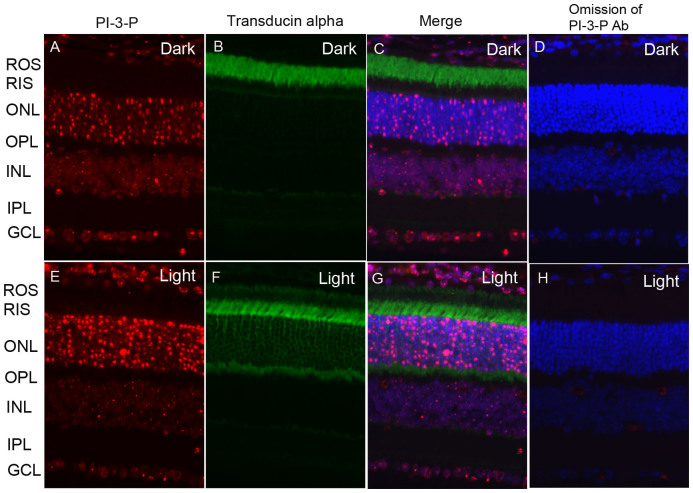
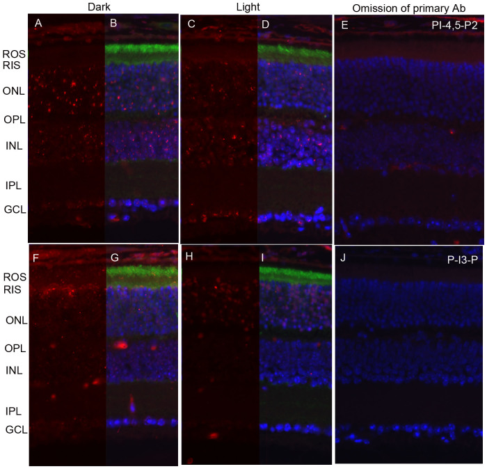
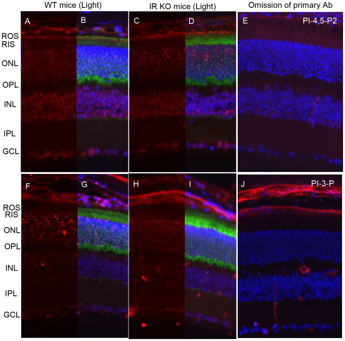
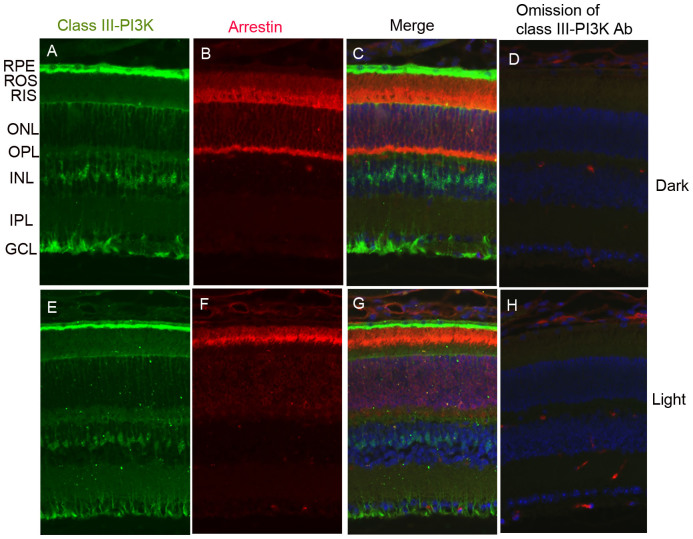
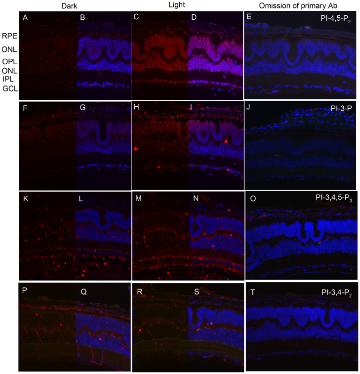
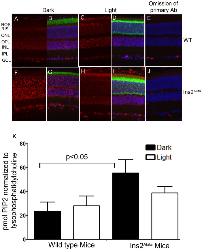
Activation of the phosphoinositide (PI) cycle generates the second messengers that control various aspects of cellular signaling. We have previously shown that two PI cycle enzymes, type II phosphatidylinositol 5-phosphate 4-kinase (PIPK IIα) and phosphoinositide 3-kinase (PI3K), are activated through light stimulation. In our earlier studies, we measured enzyme activities, instead of directly measuring the products, due to lack of sensitive analytical techniques. Cells have very low levels of PIs, compared to other lipids, so special techniques and sensitive analytical instruments are necessary for their identification and quantification. There are also other considerations, such as different responses in different cell types, which may complicate quantification of PIs. For example, although light activated PIPK IIα, there was no increase in PI-4,5-P2 measured by liquid chromatography-mass spectrometry (LC/MS) This discrepancy is due to the heterogeneous nature of the retina, which is composed of various cell types. In this study, we examined PI generation in situ using immunohistochemistry with specific PI antibodies. PIs were generated in specific retinal cell layers, suggesting that analyzing PIs from the total retina by LC/MS underscores the significance. This suggests that PI-specific antibodies are useful tools to study the cell-specific regulation of PIs in the retina.


Figures

Figure 1
Figure 1. Immunofluorescence analysis of PI-4,5-P2 in mouse retina.
Prefer-fixed sections of dark- (A–D) and light-adapted (E–H) mouse retinas were stained for PI-4,5-P2 (A, E), transducin alpha (B, F), and DAPI (C, G). Immunofluorescence was analyzed by epifluorescence. Panels C and G represent the merged images of PI-4,5-P2 and transducin alpha. Panels D and H represent the omission of PI-4,5-P2 and transducin alpha antibodies. ROS, rod outer segments; RIS, rod inner segments; ONL, outer nuclear layer; OPL, outer plexiform layer; INL, inner nuclear layer; IPL, inner plexiform layer; GCL, ganglion cell layer.
Figure 2
Figure 2. Immunofluorescence analysis of PI-3-P in mouse retina.
Prefer-fixed sections of dark- (A–D) and light-adapted (E–H) mouse retinas were stained for PI-3-P (A, E), transducin alpha (B, F), and DAPI (C, G). Immunofluorescence was analyzed by epifluorescence. Panels C and G represent the merged images of PI-3-P and transducin alpha. Panels D and H represent the omission of PI-3-P antibody. ROS, rod outer segments; RIS, rod inner segments; ONL, outer nuclear layer; OPL, outer plexiform layer; INL, inner nuclear layer; IPL, inner plexiform layer; GCL, ganglion cell layer.
Figure 3
Figure 3. Generation of PI-4,5-P2 and PI-3-P is under the control G-protein coupled receptor rhodopsin activation.
Prefer-fixed sections of dark- (A–E) and light-adapted (F–J) Rpe65-/- mouse retinas were stained for PI-4,5-P2 (A, C), PI-3-P (F, H), transducin alpha (B, D, G, I), and DAPI (B, D, G, I). Immunofluorescence was analyzed by epifluorescence. Panels B, D, G, and I represent the merged images of either PI-4,5-P2 or PI-3-P with transducin alpha. Panels E and J represent the omission of primary antibodies. ROS, rod outer segments; RIS, rod inner segments; ONL, outer nuclear layer; OPL, outer plexiform layer; INL, inner nuclear layer; IPL, inner plexiform layer; GCL, ganglion cell layer.
Figure 4
Figure 4. Generation of PI-3-P is under the control of insulin receptor activation.
Prefer-fixed sections of light-adapted wild type (A, B, F, G) and IR KO (C, D, H, I) mouse retinas were stained for PI-4,5-P2 (A–D), PI-3-P (F–I), transducin alpha, and DAPI (B, D, G, J). Immunofluorescence was analyzed by epifluorescence. Panels B, D, G, and I represent the merged images of either PI-4,5-P2 or PI-3-P with transducin alpha. Panels E and J represent the omission of primary antibodies. ROS, rod outer segments; RIS, rod inner segments; ONL, outer nuclear layer; OPL, outer plexiform layer; INL, inner nuclear layer; IPL, inner plexiform layer; GCL, ganglion cell layer.
Figure 5
Figure 5. Immunofluorescence analysis of class III PI3K in mouse retina.
Prefer-fixed sections of dark- (A–D) and light-adapted (E–H) mouse retinas were stained for class III PI3K (A, E), arrestin (B, F), and DAPI (C, G). Immunofluorescence was analyzed by epifluorescence. Panels C and G represent the merged images of class III PI3K and arrestin. Panels D and H represent the omission of class III PI3K antibody. RPE, retinal pigment epithelium; ROS, rod outer segments; RIS, rod inner segments; ONL, outer nuclear layer; OPL, outer plexiform layer; INL, inner nuclear layer; IPL, inner plexiform layer; GCL, ganglion cell layer.
Figure 6
Figure 6. Immunofluorescence analysis of PI-4,5-P2, PI-3-P, PI-3,4,5-P3 and PI-3,4-P2 in cone dominant retina.
Prefer-fixed sections of dark- (A, F, K, P) and light-adapted (C, H, M, R) Nrl-/- mouse retinas were stained for PI-4,5-P2 (A, C), PI-3-P (F, H), PI-3,4,5-P3 (K, M), PI-3,4-P2 (P,R), and DAPI (B, D, G, I, L, N, Q, S). Immunofluorescence was analyzed by epifluorescence. Panels E, J, O, and T represent the omission of primary PI antibodies. RPE, retinal pigment epithelium; ROS, rod outer segments; RIS, rod inner segments; ONL, outer nuclear layer; OPL, outer plexiform layer; INL, inner nuclear layer; IPL, inner plexiform layer; GCL, ganglion cell layer.
Figure 7
Figure 7. Immunofluorescence analysis of PI-4,5-P2 in type 1 diabetic Ins2Akita mouse retina.
Prefer-fixed sections of dark- (A–E) and light-adapted (F–J) wild type (A–E) and type 1 diabetic Ins2Akita (F–J) mouse retinas were stained for PI-4,5-P2 (A, C, F, H), transducin alpha, and DAPI (B, D, G, I). Immunofluorescence was analyzed by epifluorescence. Panels B, D, G, and I represent the merged images of PI-4,5-P2 and transducin alpha. Panels E and J represent the omission of PI-4,5-P2 antibody. ROS, rod outer segments; RIS, rod inner segments; ONL, outer nuclear layer; OPL, outer plexiform layer; INL, inner nuclear layer; IPL, inner plexiform layer; GCL, ganglion cell layer. Quantification of PIP2 levels in the whole retina from dark- and light-adapted wild type and Ins2Akita mice. Polyphosphoinositides were extracted from the retina and derivatized using trimethylsilyl diazomethane, then were measured using mass spectrometry. Data are mean ± SEM, n = 6. The PIP2 levels were normalized to lysophosphatidylcholine. The significance between dark-adapted WT and type diabetic Ins2Akita shows p<0.05.

References

    1. Fruman D. A., Meyers R. E. & Cantley L. C. Phosphoinositide kinases. Annu. Rev. Biochem. 67, 481–507 (1998). - PubMed
    1. Martin T. F. Phosphoinositide lipids as signaling molecules: common themes for signal transduction, cytoskeletal regulation, and membrane trafficking. Annu. Rev. Cell Dev. Biol. 14, 231–264 (1998). - PubMed
    1. Balla T. Phosphoinositides: tiny lipids with giant impact on cell regulation. Physiol Rev. 93, 1019–1137 (2013). - PMC - PubMed
    1. Streb H., Irvine R. F., Berridge M. J. & Schulz I. Release of Ca2+ from a nonmitochondrial intracellular store in pancreatic acinar cells by inositol-1,4,5-trisphosphate. Nature 306, 67–69 (1983). - PubMed
    1. Rameh L. E. & Cantley L. C. The role of phosphoinositide 3-kinase lipid products in cell function. J. Biol. Chem. 274, 8347–8350 (1999). - PubMed

Publication types

Substances