Review
. 2014 Aug;55(8):1170-86.
doi: 10.1111/epi.12692. Epub 2014 Jun 25.

WONOEP appraisal: new genetic approaches to study epilepsy

Affiliations
Review

WONOEP appraisal: new genetic approaches to study epilepsy

Elsa Rossignol et al. Epilepsia. 2014 Aug.

Abstract

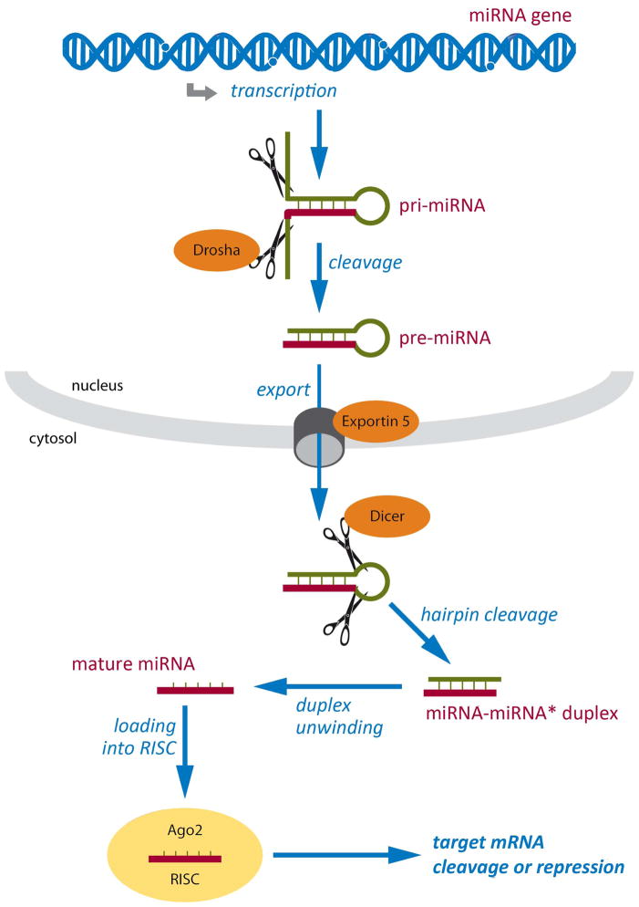
New genetic investigation techniques, including next-generation sequencing, epigenetic profiling, cell lineage mapping, targeted genetic manipulation of specific neuronal cell types, stem cell reprogramming, and optogenetic manipulations within epileptic networks are progressively unraveling the mysteries of epileptogenesis and ictogenesis. These techniques have opened new avenues to discover the molecular basis of epileptogenesis and to study the physiologic effects of mutations in epilepsy-associated genes on a multilayer level, from cells to circuits. This manuscript reviews recently published applications of these new genetic technologies in the study of epilepsy, as well as work presented by the authors at the genetic session of the XII Workshop on the Neurobiology of Epilepsy (WONOEP 2013) in Quebec, Canada. Next-generation sequencing is providing investigators with an unbiased means to assess the molecular causes of sporadic forms of epilepsy and has revealed the complexity and genetic heterogeneity of sporadic epilepsy disorders. To assess the functional impact of mutations in these newly identified genes on specific neuronal cell types during brain development, new modeling strategies in animals, including conditional genetics in mice and in utero knock-down approaches, are enabling functional validation with exquisite cell-type and temporal specificity. In addition, optogenetics, using cell-type-specific Cre recombinase driver lines, is enabling investigators to dissect networks involved in epilepsy. In addition, genetically encoded cell-type labeling is providing new means to assess the role of the nonneuronal components of epileptic networks such as glial cells. Furthermore, beyond its role in revealing coding variants involved in epileptogenesis, next-generation sequencing can be used to assess the epigenetic modifications that lead to sustained network hyperexcitability in epilepsy, including methylation changes in gene promoters and noncoding ribonucleic acid (RNA) involved in modifying gene expression following seizures. In addition, genetically based bioluminescent reporters are providing new opportunities to assess neuronal activity and neurotransmitter levels both in vitro and in vivo in the context of epilepsy. Finally, genetically rederived neurons generated from patient induced pluripotent stem cells and genetically modified zebrafish have become high-throughput means to investigate disease mechanisms and potential new therapies. Genetics has changed the field of epilepsy research considerably, and is paving the way for better diagnosis and therapies for patients with epilepsy.

Keywords: Calcium channels; Cytoskeleton; EEG monitoring; Interneurons; Systems biology; “-Omics”.


Figures

Figure 1
Figure 1. Comparative Genomic Hybridization (CGH) assays frequently reveal copy number variants (CNVs) in patients with unexplained epileptic encephalopathy
(A) Comparative Genomic Hybridization (CGH) assays are conducted by comparing a patient’s genomic DNA, labeled with a fluorescent dye such as fluorescein (green), to a control DNA, labeled with another fluorescent dye such as rhodamine (red), both applied on a microarray chip in which each well contains a probe specific for a given genomic interval. Deletions (red) and amplifications (green) in the proband’s DNA can be differentiated from areas with balanced (normal) DNA. The sensitivity of CGH analysis is inversely proportional to the spacing of consecutive probes, usually around 500Kb. (B) Examples of microdeletions (red) and microduplications (blue) presenting with neurodevelopmental phenotypes and reported in the Angelman’s syndrome interval 15q11-13, encompassing the UBE3A gene (generated using the UCSC Genome Browser on March 17th, 2014).
Figure 2
Figure 2. Whole exome sequencing often reveals detrimental de novo mutations in patients with sporadic epileptic encephalopathies
(A) Schematic representation of the procedure involved in conducting whole exome sequencing. The proband’s genomic DNA is first sheared in ≈200bp fragments which are protected with end-adaptors. Secondly, the patient’s DNA is hybridized to an exome capture library consisting of specific probes designed to recognize most coding fragments of human DNA (i.e. exons and adjacent intron-exon splice-site junctions). The hybridized fragments are extracted using systems such as streptavidin-labeled beads. The patient’s exonic DNA is then retrieved and sent for sequencing (massive parallel sequencing). (B) The sequences obtained are aligned to the reference human genome sequence. Multiple reads will be obtained for each genomic interval sequenced. De novo variants that are unique to the proband and not inherited from the parents can be identified. These variants are then confirmed using Sanger re-sequencing (illustrated in bottom panel). In this particular patient presenting with early-onset epileptic encephalopathy, exome sequencing revealed a single de novo variant, where an A replaces the reference G in a heterozygous fashion (c. G875A), in the well-known epileptic encephalopathy gene STXBP1. This variant was predicted pathogenic by different bioinformatic scores, such as SIFT and PolyPHEN, which consider the variant’s impact on protein structure and domain conservation.
Figure 3
Figure 3. Conditional genetic strategies to generate mutant mice carrying cell-type selective mutations
(A) Conditional genetic strategies allow the generation of mutant mice carrying a specific loss-of-function mutation in a gene of interest in a cell-type and tissue specific manner. Driver mouse lines are selected based on their expression of Cre recombinase driven by promoters expressed selectively in the cell-types and tissues of interest. This line is then bred on a conditional mutant mouse line carrying a floxed allele of the gene of interest, in which Lox P sites have been inserted around specific exons. The floxed allele is expressed properly in all tissues except in cells that express the Cre recombinase. In these mutated cells, the lox P sites will be recombined, effectively generating a deletion between the 2 sites, often leading to a loss-of-function allele. A conditonal reporter mouse line can be bred unto these mutants, allowing for the specific labeling of cells expressing the Cre recombinase. The mutated cells can then be tracked in a reliable fashion for their entire life-time as EGFP will be expressed stably over time. In the example illustrated here, an Nkx2.1Cre driver line was used to selectively ablate the 4th exon of the Cacna1a gene, leading to a loss-of-function allele. (B) The Nkx2.1Cre line was selected as it efficiently recombines the majority of cortical and limbic GABAergic interneurons derived form the medial ganglionic eminence, including the parvalbumin (PV) and the somatostatin (SST) expressing populations, while sparing the GABAergic cell populations in the thalamus reticular nucleus (RN). (C) Mutant cells can then be assessed using a variety of techniques, such as immunohistochemistry and in vitro physiology. In the example illustrated here, paired recordings between cortical fast-spiking (FS) GABAergic interneurons (identified using the RCEEGFP conditional labeling) and connected pyramidal cells revealed a significant alteration in the synaptic release properties of mutated FS interneurons in conditional Nkx2.1Cre; Cacna1ac/c;RCEEGFP mutant mice compared to littermate controls. These conditional mutants developed severe early-onset generalized seizures (adapted from Rossignol et al., Annals of Neurology, 2013).
Figure 4
Figure 4. Invalidating myoclonin1 disrupts radial neuronal migration
Downregulation of myoclonin1 using an shRNA delivered through in utero electroporation reveals a substantial impairement in neuronal migration and confirms the role of this gene in cortical development (CP: Cortical plate, IZ: intermediate zone, VZ/SVZ: (sub)ventricular zone).
Figure 5
Figure 5. Analyzing epigenomic signatures in epilepsy using Next Generation Sequencing
Epigenetic marks can be analyzed on a genome level using massive parallel sequencing technologies also referred to as Next Generation Sequencing (NGS). To study DNA methylation, preparation of genomic DNA is required followed by a sonication step to fragment DNA (300–400bp). Methylated DNA can then be enriched using either 5-mC specific antibodies (i.e. Methylated DNA immunoprecipitation, MeDIP) or methyl-binding domain (MBD) proteins. 5-mC capture associated NGS can identify genomic regions with medium to high 5-mC content. However, for methylation analysis down to single base pair resolution genomic bisulfite sequencing is required (not shown). Kobow and colleagues recently mapped global DNA methylation patterns in a rat model of TLE. They provide the first report of unsupervised clustering of an epigenetic mark being used in epilepsy research to separate epileptic from non-epileptic animals. NGS can also be combined with Chromatin Immunoprecipitation (ChIP) or selective preparation methods for coding and non-coding RNAs (mRNA, microRNAs, long ncRNAs) to analyze histone modifications or RNA expression patterns respectively. Currently no such epigenomic data sets are available from human or experimental epilepsy tissue (N/A).
Figure 6
Figure 6. MicroRNA biogenesis and function
miRNA genes are transcribed to generate primary miRNA (pri-miRNA) transcripts with a hairpin secondary structure. Pri-miRNAs are cleaved by a multiprotein complex that includes Drosha, generating pre-miRNAs that are exported into the cytoplasm by Exportin 5. In the cytoplasm, Dicer cleaves the hairpin loop, producing miRNA duplexes that are then unwound to yield single-strand mature miRNAs. Finally, mature miRNAs are incorporated into the RNA-induced silencing complex (RISC) in a sequence of events involving several proteins, including Argonaute proteins like Ago2. Once incorporated into RISC, miRNAs guide the complex to specific mRNAs through complementary base-pairing, leading to their cleavage or repression. Respective mechanisms appear to be active in epileptic brain tissue.
Figure 7
Figure 7. Experimental design and biosensor glutamate specificity
(A) Schematics of biosensor that used a glutamate specific oxidative enzyme reaction to detect every molecule of glutamate in extracellular space where implanted (image with permission from vendor website, Pinnacle Technologies Inc. Kansas, USA). (B) Schematic of location of stereotaxic implant to biosensor into frontal neocortex (blue); placement of EEG leads (2 recording and 1 reference) and mounting screw to anchor head mount to skull. (C) Representative recording trace of the post-experiment ex-vivo calibration of glutamate biosensor shows specificity to glutamate (arrow heads). Step readings for every 10uM glutamate (three repeats) added to media were averaged for each sensor in the study (time scale bar = 1min). (D) Sensitivity of the glutamate biosensor in-vivo was tested by injecting MK801 intra-peritoneal injection (5mg/kg), which elicited an immediate (< 1min) and significant rise in the glutamate reading in the frontal cortex, by the sensor. MK801 which is an NMDA receptor antagonist is known to induce increases in endogenous glutamate levels (Wyckhuys et al., 2013) and has been used to model various neurologic disorders, such as epilepsy, schizophrenia, and Parkinson disease where abnormal glutamate transmission is hypothesized to be involved (Roenker et al., 2012). Rapid increases in glutamate levels in the recorded trace after injection artifact (arrowhead) indicate potent NMDA receptor block and potent sensitivity of biosensor in-vivo (time scale bar = 5 min).
Figure 8
Figure 8. Imaging with in vivo bioluminescence probes reveal tissue-specific activation of candidate genes following status epilepticus
(A) Representative time course of an in vivo bioluminescence experiment of promoter activation after brain insults. Initially, the virus harboring the promoter-bioluminescence reporter construct is injected in relevant brain structures such as the hippocampus. Days later, the first in vivo bioluminescence analysis (IVIS) is performed. The insult, e.g. status epilepticus is induced. Repetitive IVIS analyses are following before the animal is sacrificed for histology. (B) Representative increased in vivo bioluminescence reflects increased candidate promoter activity after SE.

References

    1. Mulley JC, Mefford HC. Epilepsy and the new cytogenetics. Epilepsia. 2011;52:423–432. - PMC - PubMed
    1. Dibbens LM, Tarpey PS, Hynes K, et al. X-linked protocadherin 19 mutations cause female-limited epilepsy and cognitive impairment. Nat Genet. 2008;40:776–781. - PMC - PubMed
    1. Scala E, Ariani F, Mari F, et al. CDKL5/STK9 is mutated in Rett syndrome variant with infantile spasms. J Med Genet. 2005;42:103–107. - PMC - PubMed
    1. Saitsu H, Kato M, Mizuguchi T, et al. De novo mutations in the gene encoding STXBP1 (MUNC18-1) cause early infantile epileptic encephalopathy. Nat Genet. 2008;40:782–788. - PubMed
    1. Carvill GL, Regan BM, Yendle SC, et al. GRIN2A mutations cause epilepsy-aphasia spectrum disorders. Nat Genet. 2013;45:1073–1076. - PMC - PubMed

Publication types