. 2014 Sep;88(18):10488-500.
doi: 10.1128/JVI.01261-14. Epub 2014 Jun 25.

Virus-induced tubule: a vehicle for rapid spread of virions through basal lamina from midgut epithelium in the insect vector

Affiliations

Virus-induced tubule: a vehicle for rapid spread of virions through basal lamina from midgut epithelium in the insect vector

Dongsheng Jia et al. J Virol. 2014 Sep.

Abstract

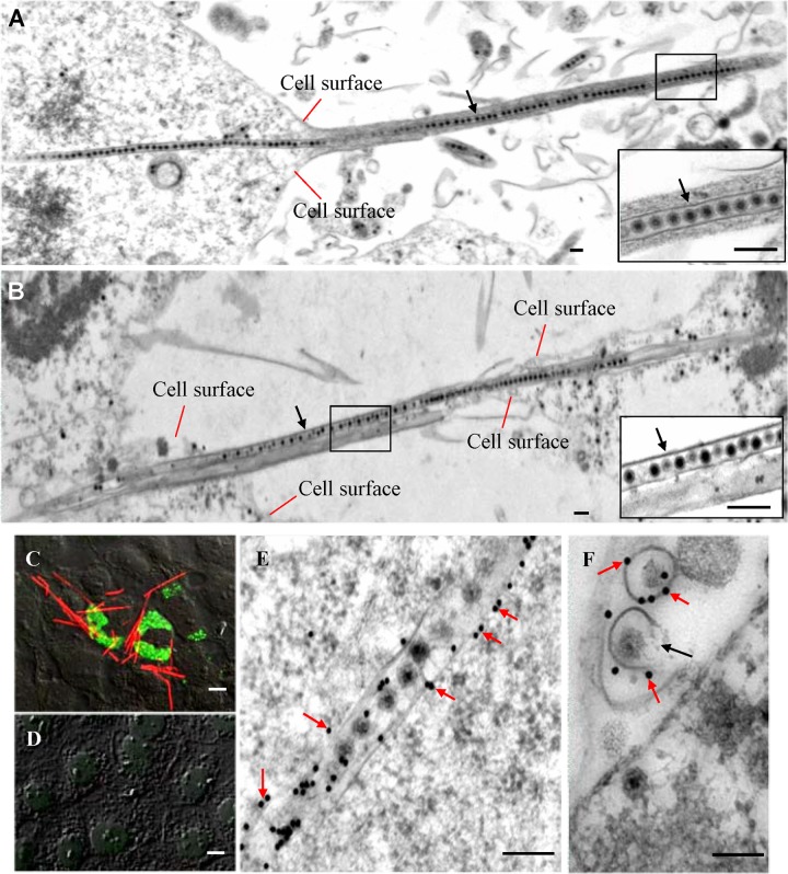
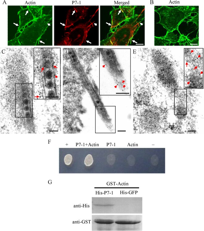
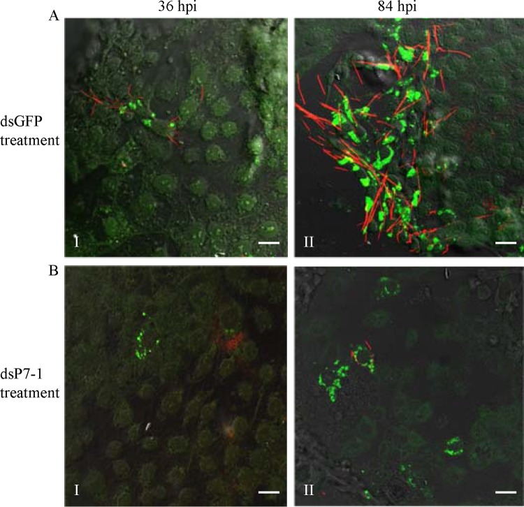
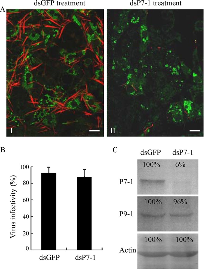
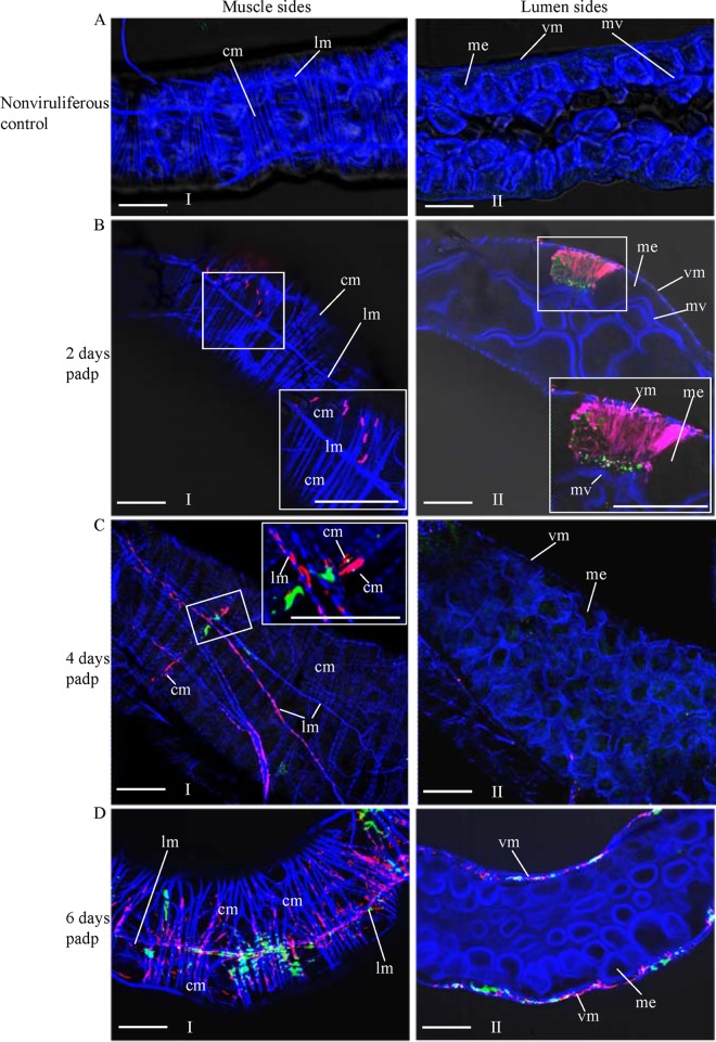
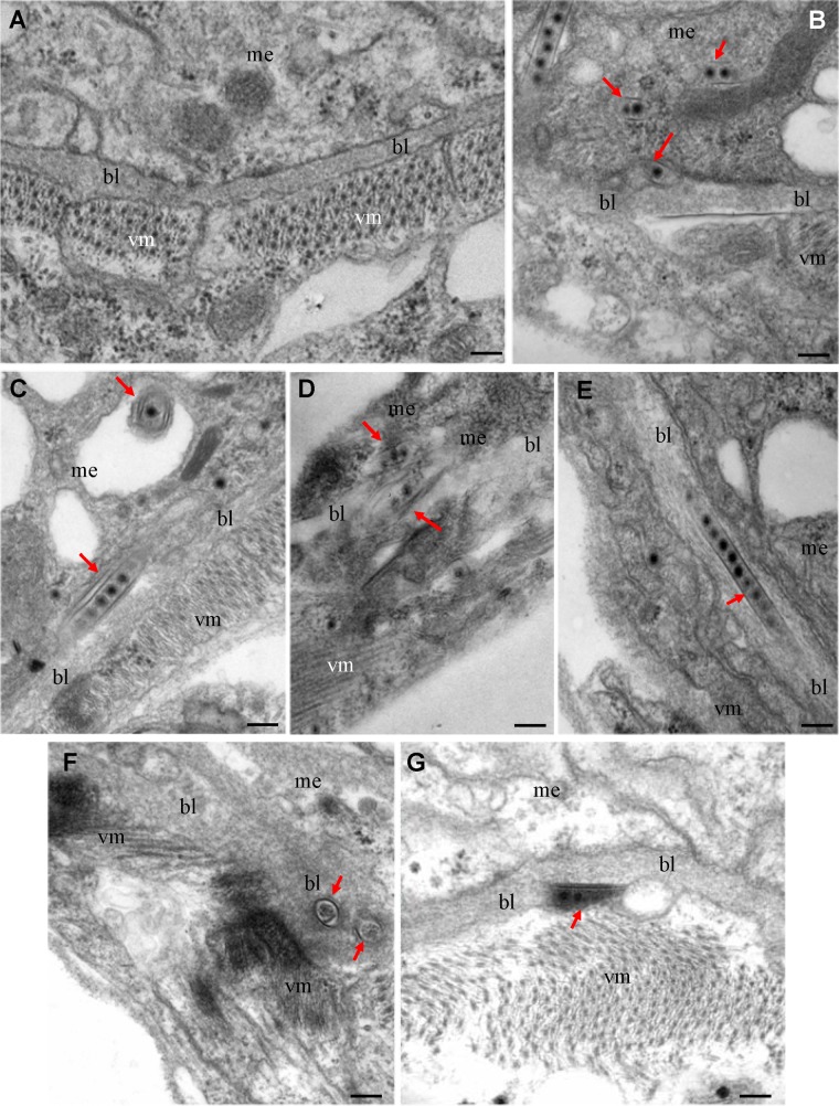
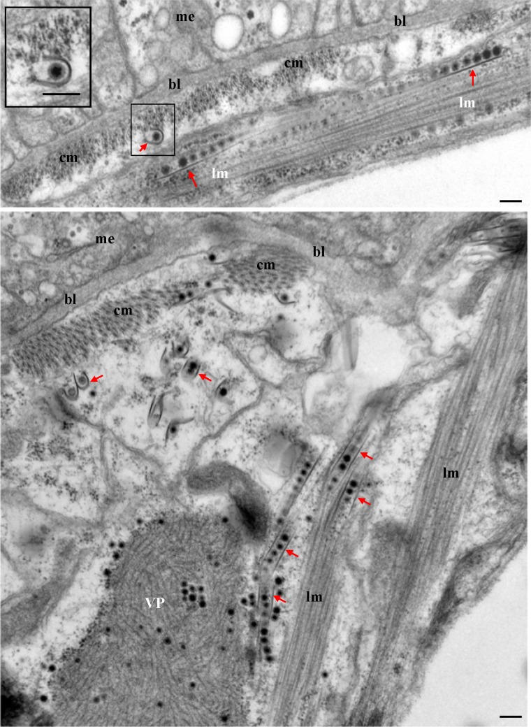
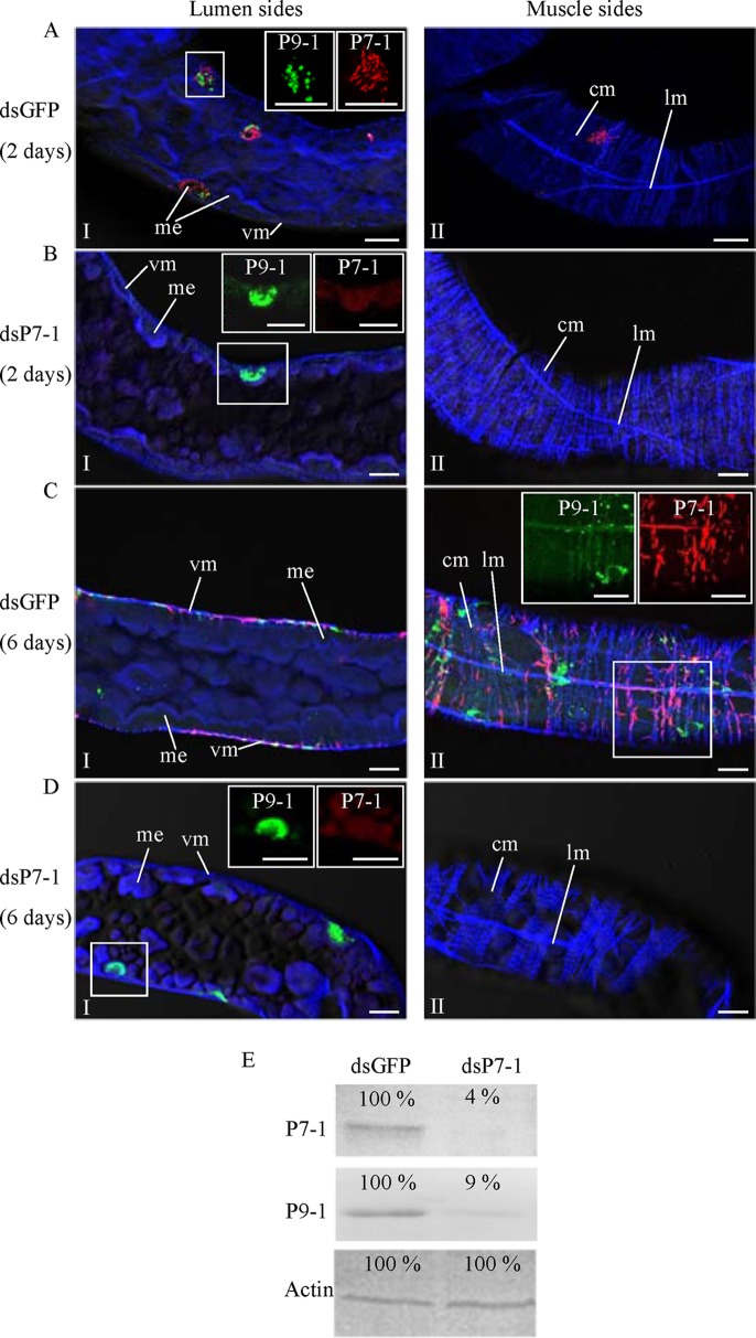
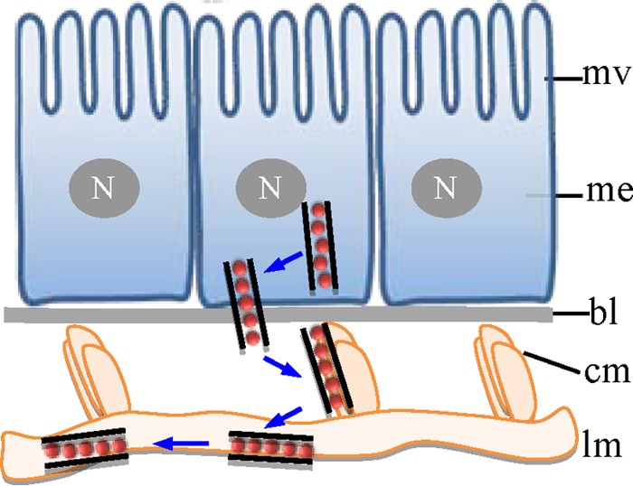
The plant reoviruses, plant rhabdoviruses, tospoviruses, and tenuiviruses are transmitted by insect vectors in a persistent propagative manner. These viruses induce the formation of viral inclusions to facilitate viral propagation in insect vectors. The intestines of insect vectors are formed by epithelial cells that lie on the noncellular basal lamina surrounded by visceral muscle tissue. Here, we demonstrate that a recently identified plant reovirus, southern rice black-streaked dwarf virus (SRBSDV), exploits virus-containing tubules composed of virus-encoded nonstructural protein P7-1 to directly cross the basal lamina from the initially infected epithelium toward visceral muscle tissues in the intestine of its vector, the white-backed planthopper (Sogatella furcifera). Furthermore, such tubules spread along visceral muscle tissues through a direct interaction of P7-1 and actin. The destruction of tubule assembly by RNA interference with synthesized double-stranded RNA targeting the P7-1 gene inhibited viral spread in the insect vector in vitro and in vivo. All these results show for the first time that a virus employs virus-induced tubule as a vehicle for viral spread from the initially infected midgut epithelium through the basal lamina, facilitating the rapid dissemination of virus from the intestine of the insect vector.

Importance: Numerous plant viruses are transmitted in a persistent manner by sap-sucking insects, including thrips, aphids, planthoppers, and leafhoppers. These viruses, ingested by the insects, establish their primary infection in the intestinal epithelium of the insect vector. Subsequently, the invading virus manages to transverse the basal lamina, a noncellular layer lining the intestine, a barrier that may theoretically hinder viral spread. The mechanism by which plant viruses cross the basal lamina is unknown. Here, we report that a plant virus has evolved to exploit virus-induced tubules to pass through the basal lamina from the initially infected midgut epithelium of the insect vector, thus revealing the previously undescribed pathway adapted by the virus for rapid dissemination of virions from the intestine of the insect vector.


Figures

FIG 1
FIG 1
Tubules were formed by nonstructural protein P7-1 of SRBSDV in virus-infected VCMs. (A, B) Electron micrographs of the virus-containing tubules (arrows) protruding from the cell surface to connect neighboring cells at 84 hpi. (Insets) Enlarged images of the boxed areas. Bars, 100 nm. (C, D) Confocal immunofluorescence micrographs of the tubules protruding from the surface of infected cells at 84 hpi. SRBSDV-infected (C) and mock-infected (D) VCMs were immunolabeled with P9-1–FITC (green) and P7-1–rhodamine (red). Bars, 5 μm. (E, F) Immunogold labeling of P7-1 of SRBSDV in the tubules in longitudinal (E) or transverse (F) sections at 84 hpi. Cells were immunolabeled with P7-1-specific IgG as the primary antibody, followed by treatment with 15-nm gold particle-conjugated goat antibodies against rabbit IgG as secondary antibodies. Black arrow in panel F, an incompletely closed tubule. Bars, 100 nm.
FIG 2
FIG 2
Association of P7-1 tubules with actin filaments in VCMs. (A, B) Immunofluorescence micrographs of the colocalization of P7-1 tubules with actin filaments (arrows) in VCMs at 84 hpi. SRBSDV-infected (A) and mock-infected (B) VCMs were immunolabeled with the actin dye FTIC-phalloidin (green) and P7-1–rhodamine (red). Bars, 5 μm. (C to E) Immunoelectron micrographs of the association of some actin filaments with virus-containing tubules within the cytoplasm (C) or along the filopodia (D) at 84 hpi. SRBSDV-infected (C, D) and mock-infected (E) VCMs were immunolabeled with actin-specific IgG as the primary antibody, followed by treatment with 10-nm gold particle-conjugated goat antibodies against rabbit IgG as secondary antibodies. (Insets) Enlarged images of the boxed areas. Arrows, gold particles. Bars, 100 nm. (F) The interaction between P7-1 of SRBSDV and actin of the WBPH detected by yeast two-hybrid assay. Transformants on an SD-Trp-Leu-His-Ade agar plate are shown. +, positive control, i.e., pBT3-STE and pOst1-NubI; P7-1+Actin, pBT3-STE-P7-1 and pPR3-N-actin; P7-1, pBT3-STE-P7-1 and pPR3-N; Actin, pBT3-STE and pPR3-N-actin; −, negative control, i.e., pBT3-STE and pPR3-N. (G) The pulldown assay was used to detect the interaction between P7-1 of SRBSDV and actin of WBPH. P7-1 of SRBSDV was fused with His to act as a bait protein with GFP as a control. Actin of WBPH was fused with GST as a prey protein. Actin bound to His-fused P7-1 of SRBSDV, but it did not bind to His-fused GFP.
FIG 3
FIG 3
RNAi induced by dsP7-1 inhibited the assembly of tubules and the spread of SRBSDV in VCMs. At 24 h after transfection with dsGFP (A) or dsP7-1 (B), VCMs were inoculated with SRBSDV at an MOI of 0.04. At 36 hpi (panels I) or 84 hpi (panels II), VCMs were immunolabeled with P9-1–FITC (green) and P7-1–rhodamine (red) and then examined by confocal microscopy. Images are representative of those from more than 4 experiments. Bars, 20 μm.
FIG 4
FIG 4
RNAi induced by dsP7-1 knocks down the expression of P7-1 without significantly inhibiting virus replication in VCMs. (A) dsP7-1 did not significantly affect the viral infection rate in VCMs. VCMs were transfected with dsGFP (panel I) or dsP7-1 (panel II) and then inoculated with SRBSDV at an MOI of 10. At 84 hpi, cells were immunolabeled with P9-1–FITC (green) and P7-1–rhodamine (red) and then examined by confocal microscopy. Images are representative of those from more than 4 experiments. Bars, 20 μm. (B) Effect of treatment with dsRNAs on the accumulation of cell-associated SRBSDV in VCMs at 84 hpi. Viral titers were determined in duplicate by a fluorescent focus assay. Error bars indicate standard deviations from three independent experiments. (C) A Western blotting assay showed that dsP7-1 significantly reduces the accumulation of P7-1 but not P9-1 of SRBSDV in virus-infected VCMs at 84 hpi. Proteins were separated by SDS-PAGE to detect P7-1 or P9-1 with P7-1- or P9-1-specific IgGs, respectively. Insect actin was detected with actin-specific IgG as a control. The protein accumulation level in VCMs transfected with dsGFP was taken to be 100%.
FIG 5
FIG 5
Extension of P7-1 tubules from the initially infected midgut epithelium toward visceral muscle tissues in viruliferous WBPHs. The internal organs of nonviruliferous WBPHs (A) and viruliferous WBPHs at 2 days (B), 4 days (C), or 6 days (D) padp were immunolabeled with P9-1–FITC (green), P7-1–rhodamine (red), and the actin dye phalloidin-Alexa Fluor 647 carboxylic acid (blue) and then examined by confocal microscopy. (Panels I and II) the muscle (I) and lumen (II) sides of the midgut. (Insets) Enlarged images of the boxed areas. Images are representative of those from more than 4 experiments. me, midgut epithelium; mv, microvilli; vm, visceral muscle; cm, circular muscle; lm, longitudinal muscle. Bars, 30 μm.
FIG 6
FIG 6
Electron micrographs showing the direct passing of virus-containing tubules through the midgut basal lamina in viruliferous WBPHs at 2 days padp. (A) The midgut of nonviruliferous WBPH; (B, C) the tubules were attached to the basal lamina in the epithelium; (D to F) the entire tubules were completely embedded in the basal lamina in longitudinal (D, E) or transverse (F) sections; (G) the tubule was closely associated with the basal lamina in the visceral muscle tissues. Arrows, virus-containing tubules. bl, basal lamina; me, midgut epithelium; vm, visceral muscle. Bars, 100 nm.
FIG 7
FIG 7
Electron micrographs showing the association of virus-containing tubules with the circular and longitudinal muscle fibers lining the midgut epithelium at 6 days padp. (Inset) Enlarged image of the boxed area. VP, viroplasm; bl, basal lamina; me, midgut epithelium; cm, circular muscle; lm, longitudinal muscle; arrows, virus-containing tubules. Bars, 100 nm.
FIG 8
FIG 8
Microinjection of dsP7-1 suppressed viral spread in vivo in WBPHs. The nymphs of WBPHs were microinjected with dsGFP (A, C) or dsP7-1 (B, D). At 2 days padp (A, B) or 6 days padp (C, D), the internal organs of WBPHs receiving dsGFP or dsP7-1 were immunolabeled with P9-1–FITC (green), P7-1–rhodamine (red), and the actin dye phalloidin-Alexa Fluor 647 carboxylic acid (blue) and then examined by confocal microscopy. (Panels I and II) The lumen (I) and muscle (II) sides of the midgut. (Insets) Green fluorescence (P9-1 antigens) and red fluorescence (P7-1 antigens) of the merged images in the boxed areas. Images are representative of those from more than 4 experiments. me, midgut epithelium; vm, visceral muscle; cm, circular muscle; lm, longitudinal muscle. Bars, 15 μm. (E) Detection of viral proteins P7-1 and P9-1 in WBPHs by Western blotting assay with P7-1-specific or P9-1-specific IgGs. Insect actin was detected with actin-specific IgG as a control. The protein accumulation level in WBPHs that received dsGFP was taken to be 100%.
FIG 9
FIG 9
Proposed model for the trafficking of P7-1 tubules in vivo in its insect vector, WBPH. Virus-containing P7-1 tubules pass across the basal lamina from the initially infected midgut epithelium, traffic along the internal circular muscle tissues, and then spread through the external longitudinal muscle tissues. bl, basal lamina; me, midgut epithelium; mv, microvilli; cm, circular muscle; lm, longitudinal muscle; N, nucleus.

Similar articles

Cited by

References

    1. Ammar ED. 1985. Internal morphology and ultrastructure of leafhoppers and planthoppers, p 121–162 In Nault LR, Rodriguez JG. (ed), Leafhoppers and planthoppers. John Wiley & Sons, Brisbane, Australia
    1. Cheung WWK, Purcell AH. 1993. Ultrastructure of the digestive system of the leafhopper Euscelidius variegatus Kirshbaum (Homoptera: Cicadellidae), with and without congenital bacterial infections. Int. J. Insect Morphol. Embryol. 22:49–61. 10.1016/0020-7322(93)90033-W - DOI
    1. Hogenhout SA, Ammar E-D, Whitfield AE, Redinbaugh MG. 2008. Insect vector interactions with persistently transmitted viruses. Annu. Rev. Phytopathol. 46:327–359. 10.1146/annurev.phyto.022508.092135 - DOI - PubMed
    1. Ammar ED, Tsai CW, Whitfield AE, Redinbaugh MG, Hogenhout SA. 2009. Cellular and molecular aspects of rhabdovirus interactions with insect and plant hosts. Annu. Rev. Entomol. 54:447–468. 10.1146/annurev.ento.54.110807.090454 - DOI - PubMed
    1. Bragard C, Caciagli P, Lemaire O, Lopez-Moya JJ, MacFarlane S, Peters D, Susi P, Torrance L. 2013. Status and prospects of plant virus control through interference with vector transmission. Annu. Rev. Phytopathol. 51:177–201. 10.1146/annurev-phyto-082712-102346 - DOI - PubMed

Publication types

Substances

LinkOut - more resources