. 2014 Sep 1;88(17):10120-33.
doi: 10.1128/JVI.01036-14. Epub 2014 Jun 25.

A novel partitivirus that confers hypovirulence on plant pathogenic fungi

Affiliations

A novel partitivirus that confers hypovirulence on plant pathogenic fungi

Xueqiong Xiao et al. J Virol. .

Abstract

Members of the family Partitiviridae have bisegmented double-stranded RNA (dsRNA) genomes and are not generally known to cause obvious symptoms in their natural hosts. An unusual partitivirus, Sclerotinia sclerotiorum partitivirus 1 (SsPV1/WF-1), conferred hypovirulence on its natural plant-pathogenic fungal host, Sclerotinia sclerotiorum strain WF-1. Cellular organelles, including mitochondria, were severely damaged. Hypovirulence and associated traits of strain WF-1 and SsPV1/WF-1 were readily cotransmitted horizontally via hyphal contact to different vegetative compatibility groups of S. sclerotiorum and interspecifically to Sclerotinia nivalis and Sclerotinia minor. S. sclerotiorum strain 1980 transfected with purified SsPV1/WF-1 virions also exhibited hypovirulence and associated traits similar to those of strain WF-1. Moreover, introduction of purified SsPV1/WF-1 virions into strain KY-1 of Botrytis cinerea also resulted in reductions in virulence and mycelial growth and, unexpectedly, enhanced conidial production. However, virus infection suppressed hyphal growth of most germinating conidia of B. cinerea and was eventually lethal to infected hyphae, since very few new colonies could develop following germ tube formation. Taken together, our results support the conclusion that SsPV1/WF-1 causes hypovirulence in Sclerotinia spp. and B. cinerea. Cryo-EM (cryo-electron microscopy) reconstruction of the SsPV1 particle shows that it has a distinct structure with similarity to the closely related partitiviruses Fusarium poae virus 1 and Penicillium stoloniferum virus F. These findings provide new insights into partitivirus biological activities and clues about molecular interactions between partitiviruses and their hosts.

Importance: Members of the Partitiviridae are believed to occur commonly in their phytopathogenic fungal and plant hosts. However, most partitiviruses examined so far appear to be associated with latent infections. Here we report a partitivirus, SsPV1/WF-1, that was isolated from a hypovirulent strain of Sclerotinia sclerotiorum and describe its biological and molecular features. We have demonstrated that SsPV1 confers hypovirulence. Furthermore, SsPV1 can infect and cause hypovirulence in Botrytis cinerea. Our study also suggests that SsPV1 has a vigorous ability to proliferate and spread via hyphal contact. SsPV1 can overcome vegetative incompatibility barriers and can be transmitted horizontally among different vegetative compatibility groups of S. sclerotiorum, even interspecifically. Cryo-EM reconstruction of SsPV1 shows that it has a distinct structure with similarity to closely related partitiviruses. Our studies exploit a novel system, SsPV1 and its hosts, which can provide the means to explore the mechanisms by which partitiviruses interact with their hosts.


Figures

FIG 1
FIG 1
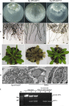
Phenotypes associated with hypovirulence of different strains of S. sclerotiorum. (A) Colony morphology of strains WF-1, Ep-1PNA367hyg, and Ep-1PNA367hygV. The latter strain represents the strain that arises from contact between Ep-1PNA367hyg and hypovirulent strain WF-1. All strains were cultured on plates with 20 ml PDAY medium at 20°C to 22°C for 3 days prior to photography. Compared to strain Ep-1PNA367hyg, strains WF-1 and Ep-1PNA367hygV exhibit low growth rates. (B) Strains WF-1 and Ep-1PNA367hygV exhibit excessive branching compared to strain Ep-1PNA367hyg. Moreover, cytoplasm was observed to extrude from some mycelial tips in the strains that contain the 2.3-kb dsRNA segments. (C) Assay of A. thaliana leaves that show hypovirulence when infected with strains WF-1 and Ep-1PNA367hygV (20°C; 4 days postinfection). (D) Ultrastructure of cells of S. sclerotiorum strain WF-1 examined by thin-section transmission electron microscopy (TEM). The ultrastructure of the hyphal tip of strain Ep-1PNA367 shows a well-distributed cytoplasm with many mitochondria whose membranes appear normal and contain many intact cristae. The ultrastructure of the hyphal tips in hypovirulent strain WF-1 shows a granulated cytoplasm and damaged mitochondria with abnormal cristae; only a few normal mitochondria remained. W, cell wall; M, mitochondria. (E) Detection of dsRNA in mycelia of strains Ep-1PNA367hyg (lanes 1 and 4), WF-1 (lanes 2 and 5), and Ep-1PNA367hygV (lanes 3 and 6). Lane M, DNA size markers.
FIG 2
FIG 2
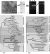
Characterization of SsPV1/WF-1 isolated from hypovirulent strain WF-1. (A) TEM images of negatively stained particles of SsPV1/WF-1. (B) SDS-PAGE of purified SsPV1/WF-1 virus particles showing one prominent band (right lane) corresponding to the viral CP. (C) Agarose gel electrophoresis of dsRNA extracted from SsPV1/WF-1 (lane 1) and mycelia of strain WF-1 (lane 2). (D) Ethidium bromide-stained, nondenaturing 15% polyacrylamide gel electrophoresis of dsRNA extracted from purified SsPV1/WF-1 (lane 1) and mycelia of strain WF-1 (lane 2). All dsRNA samples were treated with DNase I and S1 nuclease prior to electrophoresis. (E and F) Neighbor-joining phylogenetic trees constructed based on the complete amino acid sequences of the viral RdRp (E) or CP (F) of SsPV1/WF-1 and numerous other members of the family Partitiviridae. Bootstrap values (percent) obtained with 2,000 replicates are indicated on the branches, and branch lengths correspond to genetic distances; the scale bar at the bottom left corresponds to the genetic distance. Abbreviations of virus names and GenBank accession numbers are listed in Table S2 in the supplemental material.
FIG 3
FIG 3
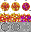
Three-dimensional structures of SsPV1/WF-1 capsids. (A to C) Surface view of three related partitiviruses, SsPV1 (A), FpV1 (B), and PsV-F (C). Two dimers in SsPV1 are highlighted in blue-shaded regions in the center. (D to F) Close-up view of the surface protrusions in SsPV1 (D), FpV1 (E), and PsV-F (F). (G to I) Central section of SsPV1 (G), FpV1 (H), and PsV-F (I) showing RNA genomes inside the particles. The capsid is radially depth cued, with the radial color bar shown on the right. The surface view of the reconstruction of SsPV1 was rendered with UCSF Chimera together with FpV1 and PsV-F.
FIG 4
FIG 4
Transfection of protoplasts of S. sclerotiorum virulent strain 1980 with purified SsPV1/WF-1 particles. (A) Comparison of colony morphologies of parental strain 1980, strain WF-1, and the SsPV1-transfected isolates 1980V1 and 1980V2. (B) Comparison of hyphal growth rates of strains 1980, WF-1, 1980V1, 1980V2, and 1980R1 (the latter is an isolate from regenerated nontransfected control protoplasts of strain 1980) on PDAY medium (20°C). (C) Numbers of sclerotia per plate produced by strains 1980, WF-1, 1980V1, 1980V2, and 1980R1 on PDAY medium (20°C; 20 days postinfection). (D) Lesion length caused by strains 1980, WF-1, 1980V1, 1980V2, and 1980R1 (20°C; 4 days postinfection) on the stems of soybean (Glycine max). (E) RT-PCR detection of SsPV1/WF-1 in individual isolates. The actin gene is used as an internal control. The predicted lengths of the RT-PCR products for CP and RdRp are 300 and 320 nt, respectively. Results in each of the histograms shown in panels B to D are expressed as arithmetic means ± standard errors of the means. Asterisks indicate a significant difference (P < 0.05) among strains of S. sclerotiorum according to the Student t test.
FIG 5
FIG 5
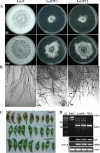
Interspecies transmission of SsPV1/WF-1 from S. sclerotiorum to S. nivalis via hyphal contact. (A) Comparison of colony morphologies of S. nivalis strain Let-19 and SsPV1-infected isolates Let-19V1 and Let-19V2, which were obtained from Let-19 after the mycelia of Let-19 contacted those of hypovirulent strain WF-1, at 5 and 10 days postinfection. (B) The hyphal tips of strains Let-19V1 and Let-19V2 showed excessive branching compared to strain Let-19. Moreover, cytoplasm (marked with black arrows) was released from some hyphal tips of strains Let-19V1 and Let-19V2. (C) Virulence assays of S. nivalis. Let-19V1 and Let-19V2 did not infect detached A. thaliana leaves at 4 days postinfection, but Let-19 did. (D) RT-PCR detection of SsPV1/WF-1 dsRNA in extracts from strain Let-19, two representative Let-19V isolates, and strain WF-1. All dsRNA samples were treated with DNase I and S1 nuclease prior to electrophoresis. The actin gene was used as an internal control. The predicted lengths of the RT-PCR products for CP and RdRp of SsPV1/WF-1 are 300 and 320 nt, respectively.
FIG 6
FIG 6
Transfection of protoplasts of virulent Botrytis cinerea strain KY-1 with purified SsPV1/WF-1 particles. (A) Comparison of colony morphologies of parental strain KY-1 and SsPV1-transfected isolates KY-1V1 and KY-1V2 at 5 and 20 days postinfection. (B) RT-PCR detection of SsPV1/WF-1 dsRNA in extracts from individual isolates. The predicted lengths of the PCR products for CP and RdRp are 300 and 320 nt, respectively. (C) Comparison of hyphal growth rates for strains KY-1, KY-1V1, and KY-1V2 on PDAY medium (20°C). (D) Comparison of numbers of sclerotia per plate produced by strains KY-1, KY-1V1, and KY-1V2 on PDAY medium (20°C; 20 days postinfection). The results plotted in each histogram are expressed as arithmetic means ± standard errors of the means. Asterisks indicate a significant difference (P < 0.05) among strains of S. sclerotiorum according to the Student t test. (E) Virulence testing on detached leaves of A. thaliana with different isolates of B. cinerea (20°C; 2 days postinfection). Strains KY-1V1 and KY-1V2 show hypovirulence. (F) Infection of B. cinerea conidia with SsPV1 is eventually lethal to infected hyphae. Plates of PDAY medium were seeded with 200-μl conidial suspensions (105 spores) of B. cinerea KY-1 and KY-1V1. Most SsPV1/WF-1-infected conidia of B. cinerea can produce a germination tube after 36 h but are unable to develop into colonies. This phenomenon was further confirmed by light microscopy.
FIG 7
FIG 7
Transmission of SsPV1/WF-1 between vegetatively incompatible host strains. (A) Colony morphology of strains R6, R6V, and 1980/R6V. R6V represents the strain that arises from R6 after contact with hypovirulent strain WF-1, whereas 1980/R6V represents strain 1980 after dual culturing and contact with strain R6V. (B) Colony morphology of strains SM-1, SM-1V, and 1980/SM-1V. SM-1V is a strain of S. minor SM-1 after strain SM-1 was dually cultured with strain WF-1 on a PDAY plate. 1980/SM-1V was a subculture that was cut out from side of strain 1980 after mycelia of strain SM-1V and 1980 contacted on a PDAY plate. (C) dsRNA extraction and RT-PCR detection of SsPV1/WF-1 in individual strains. All dsRNA samples were treated with DNase I and S1 nuclease prior to electrophoresis. The actin gene was used as an internal control. The predicted lengths of the RT-PCR products for CP and RdRp of SsPV1/WF-1 are 300 and 320 nt, whereas the predicted PCR products from SsHV1/SZ150, SsRV/SZ150, and SmRV/SM-1 are 252, 462, and 424 nt, respectively. The primers for virus detection are shown in Table S1 in the supplemental material.

Similar articles

Cited by

References

    1. Anderson PK, Cunningham AA, Patel NG, Morales FJ, Epstein PR, Daszak P. 2004. Emerging infectious diseases of plants: pathogen pollution, climate change and agrotechnology drivers. Trends Ecol. Evol. 19:535–544. 10.1016/j.tree.2004.07.021 - DOI - PubMed
    1. Strange RN, Scott PR. 2005. Plant disease: a threat to global food security. Annu. Rev. Phytopathol. 43:83–116. 10.1146/annurev.phyto.43.113004.133839 - DOI - PubMed
    1. Ghabrial SA, Suzuki N. 2009. Viruses of plant pathogenic fungi. Annu. Rev. Phytopathol. 47:353–384. 10.1146/annurev-phyto-080508-081932 - DOI - PubMed
    1. Pearson MN, Beever RE, Boine B, Arthur K. 2009. Mycoviruses of filamentous fungi and their relevance to plant pathology. Mol. Plant Pathol. 10:115–128. 10.1111/j.1364-3703.2008.00503.x - DOI - PMC - PubMed
    1. Xie J, Jiang D. New insights into mycoviruses and exploration for biological control of crop fungal diseases. Annu. Rev. Phytopathol. 10.1146/annurev-phyto-102313-050222 - DOI - PubMed

Publication types

MeSH terms

Associated data