. 2014 Sep 1;88(17):10146-56.
doi: 10.1128/JVI.01723-14. Epub 2014 Jun 25.

Structure of the herpes simplex virus 1 genome: manipulation of nicks and gaps can abrogate infectivity and alter the cellular DNA damage response

Affiliations

Structure of the herpes simplex virus 1 genome: manipulation of nicks and gaps can abrogate infectivity and alter the cellular DNA damage response

Samantha Smith et al. J Virol. .

Abstract

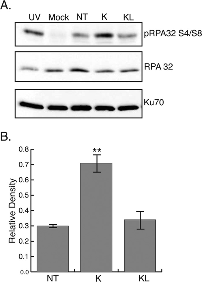
The herpes simplex virus 1 (HSV-1) virion DNA contains nicks and gaps, and in this study a novel assay for estimating the size and number of gaps in virion DNA was developed. Consistent with previous reports, we estimate that there are approximately 15 gaps per genome, and we calculate the average gap length to be approximately 30 bases. Virion DNA was isolated and treated with DNA-modifying enzymes in order to fill in the gaps and modify the ends. Interestingly, filling in gaps, blunting the ends, or adding random sequences to the 3' ends of DNA, producing 3' flaps, did not impair the infectivity of treated DNA following transfection of Vero cells. On the other hand, the formation of 5' flaps in the DNA following treatment resulted in a dramatic reduction (95 to 100%) in infectivity. Virion DNA stimulated DNA-PKcs activity in transfected cells, and DNA with 5' flaps stimulated a higher level of DNA-PKcs activity than that observed in cells transfected with untreated virion DNA. The infectivity of 5'-flapped DNA was restored in cells that do not express DNA-PKcs and in cells cotransfected with the immediate early protein ICP0, which degrades DNA-PKcs. These results are consistent with previous reports that DNA-dependent protein kinase (DNA-PK) and the nonhomologous end joining (NHEJ) repair pathway are intrinsically antiviral and that ICP0 can counteract this effect. We suggest that HSV-1 DNA with 5' flaps may induce an antiviral state due to the induction of a DNA damage response, primarily mediated by NHEJ, that renders the HSV-1 genome less efficient for lytic infection.

Importance: For productive lytic infection to occur, HSV-1 must counteract a variety of cellular intrinsic antiviral mechanisms, including the DNA damage response (DDR). DDR pathways have been associated with silencing of gene expression, cell cycle arrest, and induction of apoptosis. In addition, the fate of viral genomes is likely to play a role in whether viral genomes adopt a configuration suitable for lytic DNA replication. This study demonstrates that virion DNA activates the cellular DDR kinase, DNA-PK, and that this response is inhibitory to viral infection. Furthermore, we show that HSV-1 ubiquitin ligase, ICP0, plays an important role in counteracting the negative effects of DNA-PK activation. These findings support the notion that DNA-PK is antiviral and suggest that the fate of incoming viral DNA has important consequences for the progression of lytic infection. This study underscores the complex evolutionary relationships between HSV and its host.


Figures

FIG 1
FIG 1
Purified HSV-1 DNA contains gaps than can be filled by DNA polymerase. (A) The incorporation of labeled nucleotides by the Klenow fragment of E. coli DNA polymerase I into 200 ng of HSV-1 DNA was performed as described in Materials and Methods. For lanes 1 to 6, prior to incubation in the reaction mixture, the DNA was boiled for 2 min and quickly cooled, and random primers were added. Samples were analyzed by agarose gel electrophoresis. (B) Quantification of labeled nucleotides incorporated into the HSV-1 DNA.
FIG 2
FIG 2
Incorporation of labeled nucleotides into HSV-DNA by Klenow fragment polymerase alone, Klenow and ligase together, or T4 DNA polymerase. Purified HSV-1 DNA was incubated with labeled nucleotides and the following enzymes, as described in Materials and Methods: Klenow fragment, Klenow fragment together with T4 DNA ligase, and T4 DNA polymerase.
FIG 3
FIG 3
Virion DNA before and after treatment with Klenow or Klenow and ligase. Biotinylated nucleotides were incorporated into HSV DNA using Klenow alone or Klenow and ligase. Pulsed-field gel electrophoresis was performed with untreated (WT), Klenow-treated (K), and Klenow-ligase-treated (KL) HSV virion DNA. The gel was probed with ethidium bromide (total DNA) and CDP-Star biotin detection reagent (incorporated nucleotides).
FIG 4
FIG 4
Expected structure and measured infectivity of HSV virion DNA following various in vitro treatments. Virion DNA was treated with the indicated enzymes and tested for infectivity. The numbers of plaques have been normalized, with the plaque number obtained in control reactions set at 100%.
FIG 5
FIG 5
Transfection efficiency of treated and untreated KOS-GFP DNA. Vero cells were transfected with untreated (black), Klenow-treated (red), or Klenow-ligase-treated (blue) KOS-GFP DNA. The graph depicts overlaid histograms of cells sorted by FACS and gated for GFP-positive cells.
FIG 6
FIG 6
HSV-1 DNA stimulates RPA phosphorylation in transfected cells but not infected cells. Vero cells were either infected with HSV-1 at an MOI of 10 and harvested at 3 h postinfection (KOS) or transfected with 700 ng of virion DNA and harvested 3 h following serum addition (KOS DNA). As a positive control for pRPA32 S4/S8, Vero cells were treated with 50 J/m2 and allowed to recover for 1 h at 37°C (UV).
FIG 7
FIG 7
ICP0 prevents RPA32 S4/S8 phosphorylation in Vero cells. Vero cells were transfected with ICP0 wt or the FXE mutant and then irradiated with 100 J/m2 of UV. Cells were allowed to recover for 2 h and then fixed and stained as indicated. Arrows highlight cells transfected with ICP0.
FIG 8
FIG 8
Addition of 5′ flaps to virion DNA increases hyperphosphorylation of RPA32. (A) Vero cells were transfected with purified KOS DNA that was either left untreated (NT) or treated with Klenow (K) or Klenow and ligase (KL). Samples were harvested at 3 h following serum addition. As a positive control for pRPA S4/S8, Vero cells were treated with 50 J/m2 and allowed to recover for 1 h at 37°C (UV). (B) Densitometry analysis of Western blot in panel A. Relative density was calculated by the ratio of the densities of pRPA S4/S8 and Ku70. **, P = 0.001.
FIG 9
FIG 9
Infectivity of Klenow-treated DNA is rescued in the absence of DNA-PK. HCT-116 cells and DNA-PK−/− cells were transfected with 500 ng of untreated or Klenow-treated virion DNA. Samples were harvested at 48 h following serum addition and titrated for virus yield on Vero cells. Infectivity is reported as percentage of the untreated DNA control. Viral titers were normalized to untreated DNA control set at 100%.

References

    1. Sheldrick P, Laithier M, Lando D, Ryhiner ML. 1973. Infectious DNA from herpes simplex virus: infectivity of double-stranded and single-stranded molecules. Proc. Natl. Acad. Sci. U. S. A. 70:3621–3625. 10.1073/pnas.70.12.3621 - DOI - PMC - PubMed
    1. Jacob RJ, Roizman B. 1977. Anatomy of herpes simplex virus DNA. VIII. Properties of the replicating DNA. J. Virol. 23:394–411 - PMC - PubMed
    1. Kieff ED, Bachenheimer SL, Roizman B. 1971. Size, composition, and structure of the deoxyribonucleic acid of herpes simplex virus subtypes 1 and 2. J. Virol. 8:125–132 - PMC - PubMed
    1. Frenkel N, Roizman B. 1972. Separation of the herpesvirus deoxyribonucleic acid duplex into unique fragments and intact strand on sedimentation in alkaline gradients. J. Virol. 10:565–572 - PMC - PubMed
    1. Wilkie NM. 1973. The synthesis and substructure of herpesvirus DNA: the distribution of alkali-labile single strand interruptions in HSV-1 DNA. J. Gen. Virol. 21:453–467. 10.1099/0022-1317-21-3-453 - DOI - PubMed

Publication types

LinkOut - more resources