. 2014 Sep;88(18):10501-10.
doi: 10.1128/JVI.01347-14. Epub 2014 Jun 25.

Development of giant bacteriophage ϕKZ is independent of the host transcription apparatus

Affiliations

Development of giant bacteriophage ϕKZ is independent of the host transcription apparatus

Pieter-Jan Ceyssens et al. J Virol. 2014 Sep.

Abstract

Pseudomonas aeruginosa bacteriophage ϕKZ is the type representative of the giant phage genus, which is characterized by unusually large virions and genomes. By unraveling the transcriptional map of the ∼ 280-kb ϕKZ genome to single-nucleotide resolution, we combine 369 ϕKZ genes into 134 operons. Early transcription is initiated from highly conserved AT-rich promoters distributed across the ϕKZ genome and located on the same strand of the genome. Early transcription does not require phage or host protein synthesis. Transcription of middle and late genes is dependent on protein synthesis and mediated by poorly conserved middle and late promoters. Unique to ϕKZ is its ability to complete its infection in the absence of bacterial RNA polymerase (RNAP) enzyme activity. We propose that transcription of the ϕKZ genome is performed by the consecutive action of two ϕKZ-encoded, noncanonical multisubunit RNAPs, one of which is packed within the virion, another being the product of early genes. This unique, rifampin-resistant transcriptional machinery is conserved within the diverse giant phage genus.

Importance: The data presented in this paper offer, for the first time, insight into the complex transcriptional scheme of giant bacteriophages. We show that Pseudomonas aeruginosa giant phage ϕKZ is able to infect and lyse its host cell and produce phage progeny in the absence of functional bacterial transcriptional machinery. This unique property can be attributed to two phage-encoded putative RNAP enzymes, which contain very distant homologues of bacterial β and β'-like RNAP subunits.


Figures

FIG 1
FIG 1
Total RNA extraction from ϕKZ-infected cells. (A) Time-lapse recording of three P. aeruginosa cells infected with ϕKZ at time zero. (B) Total RNA was extracted in triplicate from 200 μl infected cells at the indicated times postinfection and quantified spectrophotometrically. (C) Transcript abundance of 5S rRNA (black) and oprL (gray) per μl extracted RNA, as determined by quantitative RT-PCR with three biological and technical replicates.
FIG 2
FIG 2
RNA-seq analysis of the ϕKZ transcriptome. Genome-wide overview of reads mapped to the sense (red) or antisense (green) strand of the ϕKZ genome at samples taken 5, 15, and 35 min after infection. Phage genes encoding structural proteins are indicated in yellow. Early, middle, and late promoters examined in more detail are indicated as red, green, and blue arrows, respectively. An example of the antisense transcription in more detail can be found in Fig. S1 in the supplemental material.
FIG 3
FIG 3
Impact of phage infection on bacterial transcriptome. Volcano plot of the P. aeruginosa transcriptome compared to the uninfected sample, with each dot representing an ORF, 10 (blue) and 35 (red) min postinfection. Triangles indicate structural RNAs, and large circles indicate genes which are located in the Pf1 operon.
FIG 4
FIG 4
Mapping the ϕKZ promoters. (A) Kinetics of accumulation of selected early, middle, and late ϕKZ transcripts, as revealed by primer extension assay, is shown. Numbers of genes located downstream of primer extension endpoints and time points when infected cells were collected and processed for RNA purification are indicated. (B) Alignments of ϕKZ promoter sequences. Consensus sequences of early, middle, and late promoters derived from 28, 6, and 16 sequences, respectively. Experimentally defined transcription start sites are in boldface. The corresponding sequence logos are depicted below the alignments. Red bars delineate conserved promoter elements.
FIG 5
FIG 5
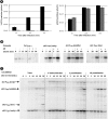
ϕKZ requires translational but not transcriptional machinery of its host. (A) Phage titers of the culture supernatant of LUZ19 and ϕKZ in the absence (black) and presence (gray) of 400 μg/ml Rif, supplied to P. aeruginosa cell cultures 5 min before the addition of phage (MOI, 1). The graphs represent the average titers obtained from three independent experiments. (B) Results of primer extension analysis of phage transcripts from early (P54), middle (P152), and late (P153) ϕKZ promoters, performed on total RNA extracted from cells infected (inf.) in the presence (+) or absence (−) of Rif. (C) Results of primer extension analysis of phage transcripts from selected early, middle, and late ϕKZ promoters on total RNA from ϕKZ-infected cells grown in the presence of Rif and chloramphenicol (Cm), added at different time points. A primer extension reaction was performed simultaneously for several phage promoters of different temporal classes, including two early promoters, P53 and P54, one middle promoter, P50, and one late promoter, P29.
FIG 6
FIG 6
β/β′ RNAP-like subunits of ϕKZ family viruses. Subunits identified as being part of the virion are colored brown. The corresponding gene numbers are shown. The position of the metal-binding catalytic motif of the β′ subunit is indicated. Introns are indicated by black triangles and arrows. Black boxes (labeled N, I, II, and III) indicate the four clusters where Rifr mutations have been identified (46). In addition to ϕKZ, included in the alignment are Cronobacter phage CR5 (NC_021531.1), Yersinia phage ϕR1-37 (NC_016163.1), Erwinia phage ϕEaH2 (NC_019929.1), Pseudomonas phage OBP (NC_016571.1), Pseudomonas phage EL (NC_007623.1), Pseudomonas phage 201ϕ2-1 (NC_010821.1), Vibrio cyclitrophicus phage JM-2012 (NC_017975.1), and Salmonella phage SPN3US (JN641803.1). More information on mutual similarities can be found in Table S4 in the supplemental material.

References

    1. Cramer P. 2002. Multisubunit RNA polymerases. Curr. Opin. Struct. Biol. 12:89–97. 10.1016/S0959-440X(02)00294-4 - DOI - PubMed
    1. Darst SA. 2001. Bacterial DNA polymerase. Curr. Opin. Struct. Biol. 11:155–162. 10.1016/S0959-440X(00)00185-8 - DOI - PubMed
    1. Lane WJ, Darst SA. 2010. Molecular evolution of multisubunit RNA polymerases: structural analysis. J. Mol. Biol. 395:686–704. 10.1016/j.jmb.2009.10.063 - DOI - PMC - PubMed
    1. Lane WJ, Darst SA. 2010. Molecular evolution of multisubunit RNA polymerases: sequence analysis. J. Mol. Biol. 395:671–685. 10.1016/j.jmb.2009.10.062 - DOI - PMC - PubMed
    1. Iyer LM, Koonin EV, Aravind L. 2003. Evolutionary connection between the catalytic subunits of DNA-dependent RNA polymerases and eukaryotic RNA-dependent RNA polymerases and the origin of RNA polymerases. BMC Struct. Biol. 28:1. 10.1186/1472-6807-3-1 - DOI - PMC - PubMed

Publication types

MeSH terms