. 2014 Sep;35(28):8092-102.
doi: 10.1016/j.biomaterials.2014.05.083. Epub 2014 Jun 23.

Creating perfused functional vascular channels using 3D bio-printing technology

Affiliations

Creating perfused functional vascular channels using 3D bio-printing technology

Vivian K Lee et al. Biomaterials. 2014 Sep.

Abstract

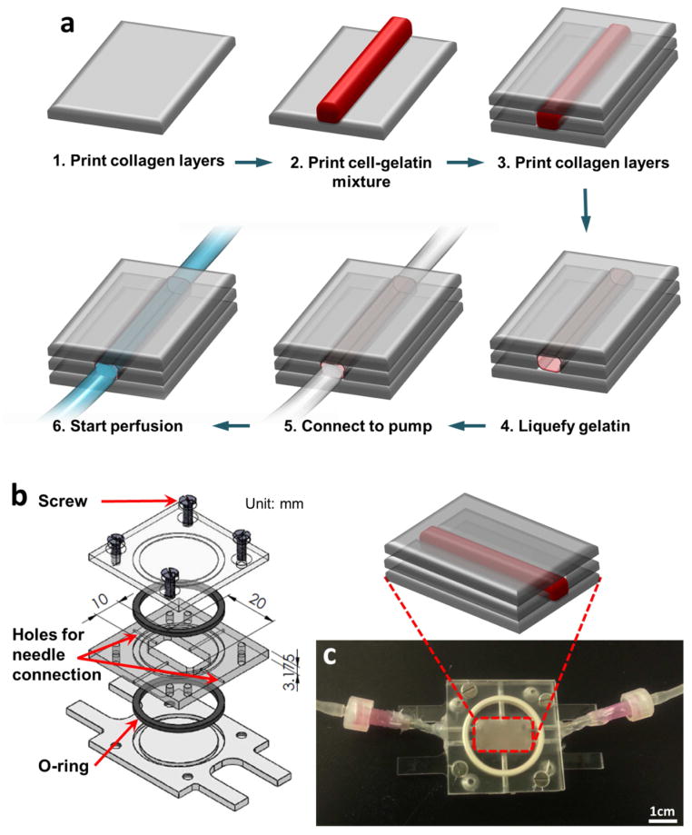
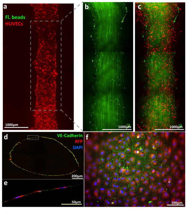
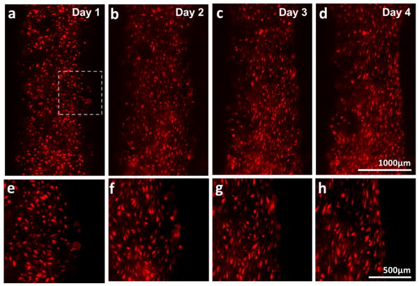
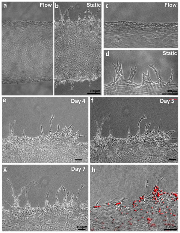
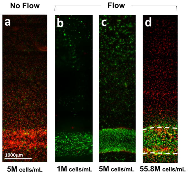
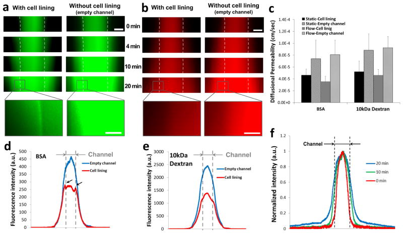
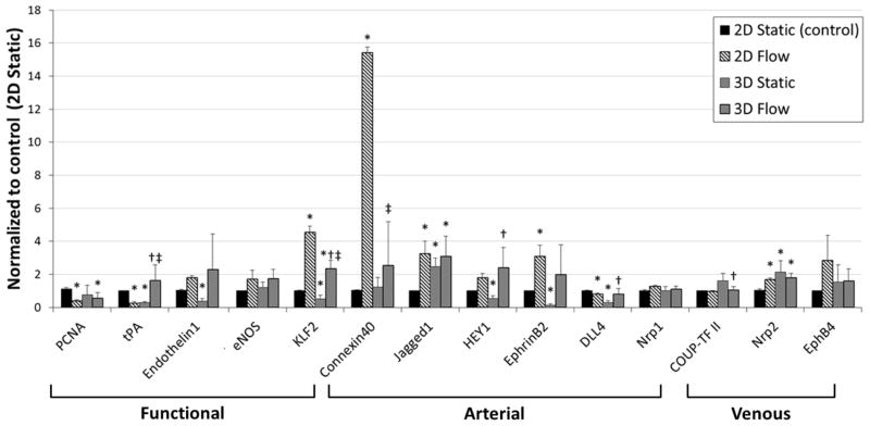
We developed a methodology using 3D bio-printing technology to create a functional in vitro vascular channel with perfused open lumen using only cells and biological matrices. The fabricated vasculature has a tight, confluent endothelium lining, presenting barrier function for both plasma protein and high-molecular weight dextran molecule. The fluidic vascular channel is capable of supporting the viability of tissue up to 5 mm in distance at 5 million cells/mL density under the physiological flow condition. In static-cultured vascular channels, active angiogenic sprouting from the vessel surface was observed whereas physiological flow strongly suppressed this process. Gene expression analysis was reported in this study to show the potential of this vessel model in vascular biology research. The methods have great potential in vascularized tissue fabrication using 3D bio-printing technology as the vascular channel is simultaneously created while cells and matrix are printed around the channel in desired 3D patterns. It can also serve as a unique experimental tool for investigating fundamental mechanisms of vascular remodeling with extracellular matrix and maturation process under 3D flow condition.

Keywords: 3D bio-printing; Hydrogel; Perfused vascularized tissue; Vascular channels.


Figures

Figure 1
Figure 1
(a) Schematics of the vascular channel construction procedure using cell-gelatin mixture. (b) Custom-designed flow chamber consists of three transparent polycarbonate pieces, two o-rings for sealing, and screws. (c) Actual picture of flow chamber. The chamber is connected to the perfusion system through needles installed on the side of chamber.
Figure 2
Figure 2
(a) Fluorescent images of vascular channel system on Day 1 of dynamic flow culture. Large image of endothelial cells (red) seeded on the printed channel. (b) Laminar flow in the vascular channel was visualized by motion of green fluorescent beads. Discontinuity of flow pattern is due to stitching (mosaic) of multiple images. (c) Overlay image of the flow of green beads and printed endothelial cells. (d) Cross-section of vascular channel after 5 days of dynamic culture. (e) magnification of dotted inset area in (d). Endothelial cells formed a monolayer along the inner surface of the channel. (f) Fluorescent image of inner surface of vascular channel after 5 days of dynamic culture. The edge of the image is out of focus due to a curved surface of the channel. (d–f) blue: DAPI nuclei staining; red: RFP-transfected HUVEC; green: VE-Cadherin.
Figure 3
Figure 3
(a–d) Alignment of HUVECs and change in channel edge shape over the dynamic culture period (Day 1 – 4). HUVECs were elongated in the direction of flow (flow from top to bottom). The edge of channel straightened. (e–h) Magnification of dotted inset area in (a–d).
Figure 4
Figure 4
Morphology of HUVECs on the vascular channel edge in dynamic culture (a, c) and static culture (b, d) on Day 5. (e–g) In the static condition, the sprouts budded from the channel edge extended over culture time, maintaining filopodia-like protrusion on the tip of the sprouts. (h) Luminal structure of sprouts was confirmed by the injection of fluorescence microbeads (10μm).
Figure 5
Figure 5
Viability assay of 3D vascular tissue. 3D vascular tissues with different cell densities were fabricated, culture for 3 days, and labeled with green (live) and red (dead) fluorescence. (a) A considerable amount of cell death was observed in the vascular tissue construct cultured in static condition (viability: < 10%). (b–c) With the media perfusion in physiological flow rate, > 90% of cells were alive in the tissue with cell density of 1 million cells/mL (b) or 5 million cells/mL density (c). (d) In a vascular tissue with cell density of 55.8 million cells/mL, cells located more than 400μm apart from channels were dead after 4 days, even with perfusion.
Figure 6
Figure 6
Time-lapse fluorescence images of (a) BSA (green color) and (b) 10kDa Dextran (red color). (c) Diffusional permeability calculation of BSA and dextran. Scale bar: 500μm, Scale bar in the magnified inset: 150 μm. (d, e) Line plots of fluorescence across the collagen scaffold and channel after 20 minutes of perfusion with (d) BSA and (e) Dextran. The position of vascular channel is indicated with grey dotted line. (f) Line plot of normalized fluorescence across the collagen scaffold and channel (indicated with black dotted line).
Figure 7
Figure 7
RNA expression of HUVECs culture in 2D static, 2D flow, 3D static, and 3D flow condition. RNA expression was measured by TaqMan RT-PCR (n = 4) on Day 5. All data are normalized to the control condition (2D Static, cultured on tissue culture plate). *p < 0.05, compared with 2D Static (control). p < 0.05, compared with 3D Static. p < 0.05, compared with 2D Flow.

References

    1. Norotte C, Marga FS, Niklason LE, Forgacs G. Scaffold-free vascular tissue engineering using bioprinting. Biomaterials. 2009;30:5910–7. - PMC - PubMed
    1. Mironov V, Visconti RP, Kasyanov V, Forgacs G, Drake CJ, Markwald RR. Organ printing: tissue spheroids as building blocks. Biomaterials. 2009;30:2164–74. - PMC - PubMed
    1. Roth EA, Xu T, Das M, Gregory C, Hickman JJ, Boland T. Inkjet printing for high-throughput cell patterning. Biomaterials. 2004;25:3707–15. - PubMed
    1. Dhariwala B, Hunt E, Boland T. Rapid prototyping of tissue-engineering constructs, using photopolymerizable hydrogels and stereolithography. Tissue Eng. 2004;10:1316–22. - PubMed
    1. Landers R, Hübner U, Schmelzeisen R, Mülhaupt R. Rapid prototyping of scaffolds derived from thermoreversible hydrogels and tailored for applications in tissue engineering. Biomaterials. 2002;23:4437–47. - PubMed

Publication types