. 2014 Nov;124(11):E437-44.
doi: 10.1002/lary.24799. Epub 2014 Jun 26.

Classification for animal vocal fold surgery: resection margins impact histological outcomes of vocal fold injury

Affiliations

Classification for animal vocal fold surgery: resection margins impact histological outcomes of vocal fold injury

Mitsuyoshi Imaizumi et al. Laryngoscope. 2014 Nov.

Abstract

Objectives/hypothesis: Extent of vocal fold injury impacts the nature and timing of wound healing and voice outcomes. However, depth and extent of the lesion created to study wound healing in animal models vary across studies, likely contributing to different outcomes. Our goal was to create a surgery classification system to enable comparison of postoperative outcomes across animal vocal fold wound-healing studies.

Study design: Prospective, controlled animal study.

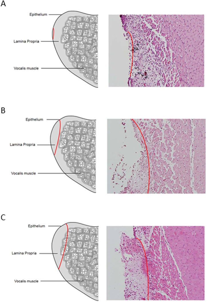
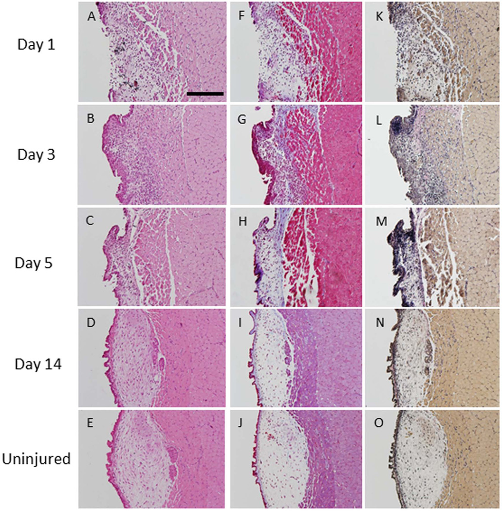
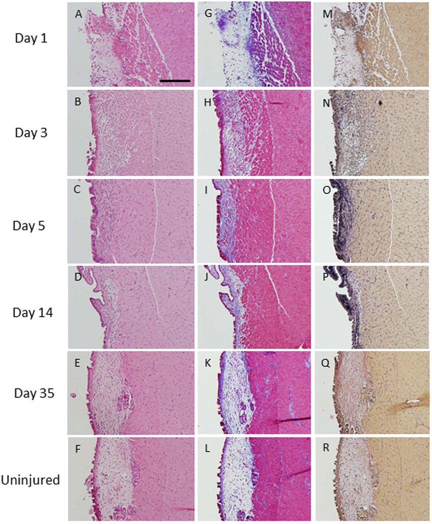
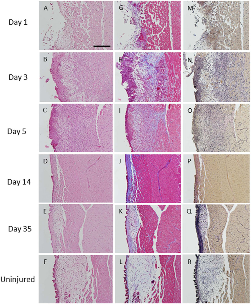
Methods: Rats underwent one of three types of unilateral vocal fold surgeries classified by depth and length of resection. The surgeries were: for subepithelial injury, resection of epithelium and superficial layer of the lamina propria at the midmembranous portion of the vocal fold; for transmucosal injury, resection of epithelium and lamina propria; and for transmuscular injury, resection of epithelium, lamina propria, and superficial portion of the vocalis muscle. Wound healing was evaluated histologically at various time points up to 35 days postinjury.

Results: Complete healing occurred by 14 days postsurgery for subepithelial injury, and by day 35 for transmucosal injury. Injury remained present at day 35 for transmuscular injury.

Conclusions: Timing and completeness of healing varied by extent and depth of resection. Scarless healing occurred rapidly following subepithelial injury, whereas scarring was observed at 5 weeks after transmuscular injury. The proposed classification system may facilitate comparison of surgical outcomes across vocal fold wound-healing studies.

Levels of evidence: N/A.

Keywords: Surgical classification; animal model; vocal fold injury; wound healing.


Conflict of interest statement

No author declares a conflict of interest.

Figures

Figure 1
Figure 1. Classification system for rat injury
Sketch and hematoxylin and eosin staining of three proposed injury types. The red line indicates the depth and extent of injury which ranges from A. resection of the epithelium and superficial layer of the lamina propria; transmucosal injury, B. resection of the epithelium and all layers of the lamina propria, and C. resection of the vocal fold mucosa and the superficial aspect of the vocalis muscle.
Figure 2
Figure 2. Subepithelial injury
Hematoxylin and eosin staining at three time points post-injury (A–D) and in an uninjured vocal fold (E). Trichrome staining shows increased collagen density (blue) up to 5 days post-injury (F–H). Collagen deposition at day 14 (I) were indistinguishable from levels observed in uninjured tissue (J). Verhoff–van Gieson (VVG) staining showed increased elastin deposition (black) in the injured lamina propria up to 5 days post-injury (K-M) compared to uninjured vocal folds (O). At day 14 (N), elastin deposition was at uninjured levels (O) (200µm scale bar; original magnification 20×).
Figure 3
Figure 3. Transmucosal injury
At day 1, epithelium was absent and inflammatory cells were observed in the lamina propria (A). The epithelium regenerated but inflammatory cells remained in the lamina propria at day 3 (B). At days 5 and 14, epithelial hyperplasia was noted and the volume of the lamina propria was reduced (C, D). The injured vocal fold closely resembled the non-injured vocal fold by day 35 (E, F). Trichrome staining showed elevated collagen deposition (blue s) at days 3, 5 and 14 (H–J). Collagen returned to baseline levels by day 35 (K, L). Elastin deposition (black) was elevated at days 3, 5 and 14 (N–P). Elastin returned to non-injured levels by day 35 (Q, R) (200 µm scale bar; original magnification 20×).
Figure 4
Figure 4. Transmuscular injury
At day 1, most of the epithelial layer and lamina propria were absent and the superficial layer of the thyroarytenoid muscle was damaged (A). Epithelial hyperplasia and infiltration of the lamina propria by inflammatory cells were observed at day 3 (B). Granulation tissue was present at day 5 (C). At days 14 and 35, a reduction in lamina propria volume and thickening of the epithelial layer was observed (D, E). Collagen density was elevated at all time points post-injury (H–K) compared to uninjured vocal folds (L). Elastin deposition was also elevated above non-injured levels at all time points (N–Q) (200µm scale bar; original magnification 20×).

References

    1. Tateya T, Tateya I, Sohn JH, Bless DM. Histologic characterization of rat vocal fold scarring. Ann Otol Rhinol Laryngol. 2005;114:183–191. - PubMed
    1. Johnson BQ, Fox R, Chen X, Thibeault S. Tissue regeneration of the vocal fold using bone marrow mesenchymal stem cells and synthetic extracellular matrix injections in rats. Laryngoscope. 2010;120:537–545. - PMC - PubMed
    1. Rousseau B, Hirano S, Chan RW, Welham N, Thibeault S, Ford C, Bless D. Characterization of chronic vocal fold scarring in a rabbit model. J Voice. 2004;18:116–124. - PubMed
    1. Thibeault SL, Klemuk SA, Chen X, Quinchia Johnson BH. In Vivo engineering of the vocal fold ECM with injectable HA hydrogels-late effects on tissue repair and biomechanics in a rabbit model. J Voice. 2011;25:249–253. - PMC - PubMed
    1. Cho SH, Kim HT, Lee IJ, Kim MS, Park HJ. Influence of phonation on basement membrane zone recovery after phonomicrosurgery: a canine model. Ann Otol Rhinol Laryngol. 2000;109:658–666. - PubMed

Publication types

LinkOut - more resources