. 2014 Aug 15;25(16):2351-64.
doi: 10.1091/mbc.E14-04-0929. Epub 2014 Jun 25.

ROCC, a conserved region in cohesin's Mcd1 subunit, is essential for the proper regulation of the maintenance of cohesion and establishment of condensation

Affiliations

ROCC, a conserved region in cohesin's Mcd1 subunit, is essential for the proper regulation of the maintenance of cohesion and establishment of condensation

Thomas Eng et al. Mol Biol Cell. .

Abstract

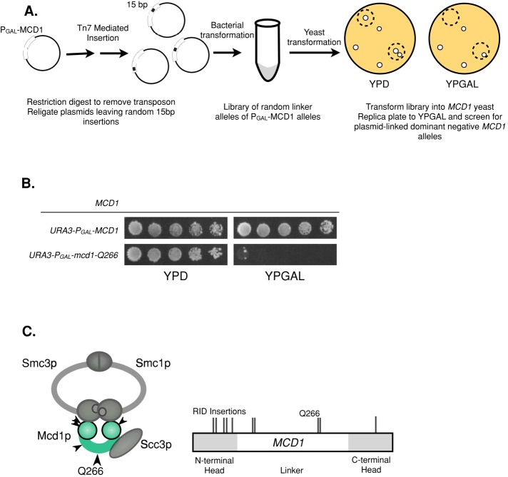
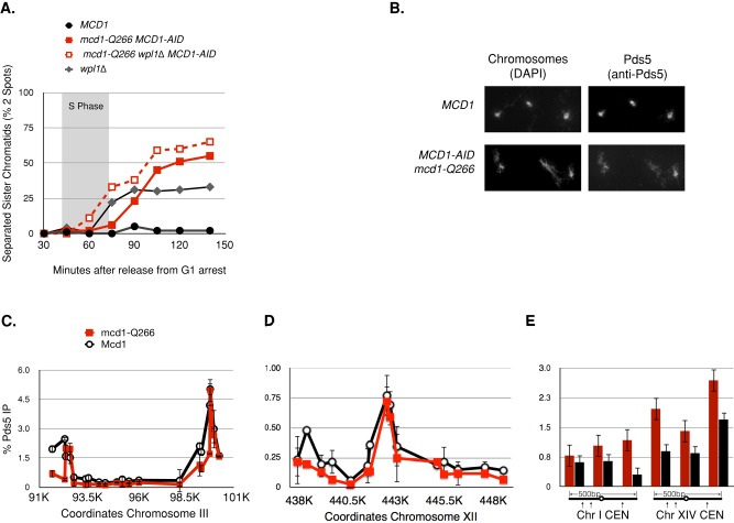
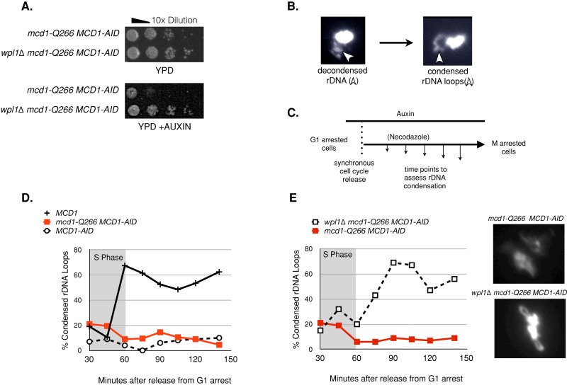
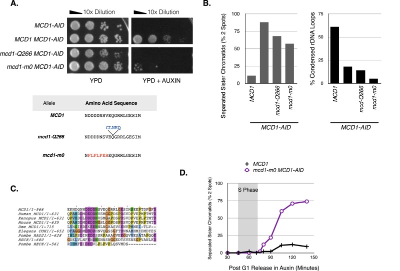
Cohesin helps orchestrate higher-order chromosome structure, thereby promoting sister chromatid cohesion, chromosome condensation, DNA repair, and transcriptional regulation. To elucidate how cohesin facilitates these diverse processes, we mutagenized Mcd1p, the kleisin regulatory subunit of budding yeast cohesin. In the linker region of Mcd1p, we identified a novel evolutionarily conserved 10-amino acid cluster, termed the regulation of cohesion and condensation (ROCC) box. We show that ROCC promotes cohesion maintenance by protecting a second activity of cohesin that is distinct from its stable binding to chromosomes. The existence of this second activity is incompatible with the simple embrace mechanism of cohesion. In addition, we show that the ROCC box is required for the establishment of condensation. We provide evidence that ROCC controls cohesion maintenance and condensation establishment through differential functional interactions with Pds5p and Wpl1p.


Figures

FIGURE 1:
FIGURE 1:
Isolation of RID mutants in MCD1. (A) Schematic of RID mutagenesis. Plasmid pVG385 containing MCD1 driven by the GAL1 promoter (pGAL-MCD1) was subjected to Tn7 transposition in vitro. The inserted transposon was removed by PmeI digestion, leaving random 15–base pair insertions. The library was transformed into either haploid WT (VG3349-1B) or mcd1-1 (VG3312-7A) strains, and transformants were screened for inviability or slow growth on galactose. (B) Haploid WT (VG3349-1B) yeast bearing pGAL-MCD1 or pGAL-mcd1-Q266 were grown at 23°C to saturation and then plated in 10-fold serial dilutions onto medium containing galactose (YPGAL) or dextrose (YPD) and incubated at 23°C for 3 d. pGAL-Mcd1-Q266 is semidominant over MCD1 and more penetrant at 16°C (unpublished data). (C) Schematic of cohesin structure and map of RID mutation positions in MCD1. The four subunits of the cohesin complex are represented. The head domains of Smc1p and Smc3p are shown as large gray balls at the base of these proteins. The amino and carboxy globular domains of Mcd1p are shown as green balls to the left and right, with the intervening green bar representing the linker region. Map of Rid mutations is shown to the right. Gray areas indicate globular domains as determined by sequence conservation.
FIGURE 2:
FIGURE 2:
mcd1-Q266 is recessive to MCD1 but exhibits a defect in cohesion maintenance after Mcd1p is removed via the auxin degron system. (A) Haploid yeast strains MCD1 (DK5531), MCD1-AID (DK5501), and mcd1-Q266 MCD1-AID (DK5535) were grown at 23°C to saturation, plated in 10-fold serial dilutions onto rich medium (YPD) or rich medium plus 500 μM auxin (YPD + auxin), and then incubated for 3 d at 23°C. (B) Schematic to measure cohesion establishment and maintenance as cells progress from G1 to M. Early-log-phase cells growing at 23°C in YPD were arrested in G1 using αF, and then auxin (500 μM) was added and cells incubated for an additional 1 h in G1. Cells were then released from G1 arrest into YPD medium containing auxin (500 μM) and nocodazole (15 μg/ml) to allow cell-cycle progression until arrest in M phase. Cell aliquots were fixed and processed every 15 min to assess cohesion and DNA content (see Supplemental Figure S3 and Material and Methods). (C) Analysis of cohesion in mcd1-Q266 cells during progression from G1 to M. Haploid MCD1 (DK5531, solid black crosses), PDS5-AID (DK5540, gray circles), MCD1-AID (DK5542, open diamonds), and mcd1-Q266 MCD1-AID (DK5535, red squares) were assessed for cohesion as described in B. The percentage of cells with two GFP foci at a CEN-distal locus (LYS4) is plotted. From 200 to 300 cells were counted at each time point. S phase is marked with a gray box (Supplemental Figure S3). The experiment was repeated three times, and representative time course is shown. (D) Schematic to measure cohesion maintenance in M phase–arrested cells. Early-log-phase cells growing at 23°C in YPD were arrested by addition of nocodazole (15 μg/ml) and incubation for 2.5 h. Auxin (500 μM) was added and cells incubated an additional 90 min. Cell aliquots were processed for the GFP cohesion assay before auxin addition (T = 0) and at 15-min intervals after auxin addition. (E) Analysis of cohesion in mcd1-Q266 strains in M-phase arrest. Haploid MCD1 (DK5531, solid black crosses), PDS5-AID (DK5540, gray circles), MCD1-AID (DK5542, open diamonds), and mcd1-Q266 MCD1-AID (DK5535, red squares) were treated and assessed for cohesion as described in D. Percentage of cells with two GFP foci at a CEN-distal locus LYS4. From 100 to 300 cells were counted at each time point. Data were generated from two independent experiments; error bars show SD.
FIGURE 3:
FIGURE 3:
mcd1-Q266p and Mcd1p exhibit similar and robust binding to chromosomes. (A) Schematic for analysis of cohesin binding as determined by chromatin immunoprecipitation. Early-log-phase cells were arrested in G1, treated with auxin to deplete MCD1-AID, and released from G1 and rearrested in M phase using nocodazole at 23°C as described in Figure 2, A and B. Cells were processed for chromosome spreads (B) and for chromatin immunoprecipitation (C–E). For ChIP experiments, the average cohesin binding from two biological replicates is plotted. Error bars represent SD between both experiments. (B) Chromosome spreads. Mcd1-3FLAG MCD1-AID (DK5536; top) and mcd1-Q266-3FLAG MCD1-AID (DK5535; bottom) cells were processed for chromosome spreads (see Materials and Methods). MCD1-Q266-3FLAGp or MCD1-3FLAGp was visualized using mouse anti-FLAG antibody (anti-FLAG) and DNA (DAPI). Data are from one of three independent experiments. (C–E) ChIP. Strains in B were processed for ChIP to assess FLAG tagged Mcd1p binding using rabbit anti-Mcd1p antibodies (Materials and Methods). Mcd1p (black lines or black bars) or mcd1-Q266p (red lines or red bars). (C) Chromosome III pericentric domain ChIP. Mcd1p binding was assessed by quantitative PCR (qPCR) using primers spanning a 10-kb region. Primer pairs were spaced every ∼500 base pairs. (D) Chromosome XII ChIP at a single-copy domain near the rDNA. Mcd1p binding over a 10-kb region was assessed by qPCR using primer pairs spaced every ∼500 base pairs. (E) CEN1 and CEN14 ChIP. Mcd1p binding at three loci immediately flanking CEN1 and CEN14. All primer sets examine DNA sequences within 500 base pairs of either centromere.
FIGURE 4:
FIGURE 4:
mcd1-Q266 and wild-type cohesin are stably bound on chromosomes at CAR sites but equally unstable at CENs. (A) Schematic to assess stability of the cohesin complex on chromosomes in M-phase cells. Cells were arrested in M phase as described in the Figure 2D legend. The culture was split in half, and auxin (500 μM) was added to one half; each half was incubated for 1 h in M phase, and then samples were processed for ChIP using rabbit anti-Mcd1 antibody (see Materials and Methods). (B, C) Analysis of cohesin binding to chromosomes in M phase after Scc2p loader depletion (stability strains). We used two haploid MCD1 stability strains, which contained (SCC2-AID MCD1-AID) and either MCD1-3FLAG (DK5560) or mcd1-Q266-3FLAG (DK5539). Strains were grown and processed as in A. FLAG-tagged Mcd1p binding at specific chromosomal sites was assessed by ChIP using qPCR. Two independent experiments were performed and gave equivalent results, one of which is shown here. (B) Effect of loader depletion on Mcd1-3FLAGp cohesin binding to chromosomes. ChIP of Mcd1-3FLAGp stability strain DK5560 when Scc2p was active (No Auxin; solid line) or after Scc2p loader was depleted (+ Auxin, dotted black line). Chip analysis at chromosome III pericentric region (left), chromosome XII single-copy arm region (middle). and within 500 base pairs of CEN3 and CEN14 (right). Data are the average of two qPCR replicates for each primer pair. (C) Effect of loader depletion on mcd1-Q266p cohesin binding to chromosomes. ChIP of Mcd1-Q266-3FLAG stability strain DK5539 when Scc2p was active (no auxin, solid red line) or after Scc2p loader was depleted (+ Auxin, dotted red line). ChIP analysis was performed at same loci as in B.
FIGURE 5:
FIGURE 5:
Mechanism of cohesion maintenance. (A) Effect of wpl1∆ on the cohesion maintenance defect of mcd1-Q266. Four haploid strains were subjected to auxin depletion in G1 phase then allowed to progress into M phase arrest under auxin depletion as described in the Figure 2B legend to assess whether WPL1 affects the mcd1-Q266 cohesion defect. Cohesion loss in MCD1 (DK5531, solid black circles), wpl1∆ (DK5561, gray diamonds), mcd1-Q266 MCD1-AID (DK5530, solid red squares), and wpl1∆ mcd1-Q266 MCD1-AID (DK5535, dotted red squares) was assessed by plotting the percentage of cells with two GFP foci at a CEN-distal locus (LYS4). From 200 to 300 cells were counted at each time point. Data are from two independent experiments. S phase is marked with a gray box. (B–E). Effect of mcd1-Q266 on Pds5p binding to chromosomes. Haploids MCD1 (DK5531) and mcd1-Q266 MCD1-AID (DK5501; bottom) were grown as described in A and then processed for chromosome spreads (B) and ChIP (C–E). (B) Chromosome spreads to detect Pds5p. The Pds5p staining in MCD1 cells (top) and mcd1-Q266 cells (bottom). Pds5p was detected using rabbit anti-Pds5p antibodies (right) and DNA detected using DAPI (left). Data were generated from two independent experiments, which gave similar results. A representative field is shown. (C–E) Pds5p ChIP. Strains in B were processed for ChIP to assess Pds5p binding using rabbit anti-Pds5p antibodies (Materials and Methods). Strains containing WT Mcd1p cohesin (DK5531, black lines) or mcd1-Q266 cohesin (DK5535, red lines) as shown. (C) Chip analysis at chromosome III pericentric region. (D) ChIP at chromosome XII single-copy arm region (middle) and (E) ChIP within 500 base pairs of CEN3 and CEN14 (right). Data from average cohesin binding from two biological replicates. Error bars represent SD between both experiments.
FIGURE 6:
FIGURE 6:
WPL1 deletion suppresses both the inviability and condensation defects of mcd1-Q266. (A) Haploid yeast strains mcd1-Q266 MCD1-AID (DK5530) and wpl1∆ mcd1-Q266 MCD1-AID (DK5550) were grown at 23°C and plated in 10-fold serial dilutions onto rich media (YPD) or rich media with 500 μM auxin (YPD + auxin). (B) Effect of wpl1∆ on the cohesion maintenance defect of mcd1-Q266. Four haploid strains were subjected to auxin depletion in G1 phase and then allowed to progress into M-phase arrest under auxin depletion as described in the Figure 2B legend. Cohesion loss in MCD1 (DK5531, solid black circles), wpl1∆ (DK5561, gray diamonds), mcd1-Q266 MCD1-AID (DK5530, solid red squares), and wpl1∆ mcd1-Q266 MCD1-AID (DK5535, dotted red squares) was assessed by plotting the percentage of cells with two GFP foci at a CEN-distal locus (LYS4). S phase is marked with a gray box. From 200 to 300 cells were counted at each time point. A representative time course is shown from two independent biological replicates. (B) Representative cytological morphology of the rDNA locus, as prepared by chromosome spreads and stained with DAPI (see Materials and Methods; Lavoie et al., 2000). Decondensed rDNA (left), condensed rDNA (right). Arrow indicates the location of rDNA. (C–F) Assessment of chromosome condensation in budding yeast. Samples were collected at 15-min intervals and processed to examine rDNA morphology by chromosome spreads stained with DAPI. At least 100 chromosome masses were scored per time point. (C) Schematic of auxin depletion in G1 and subsequent release to M-phase arrest. (D) Effect of mcd1-Q266 on rDNA condensation as described in C. Haploids MCD1 (DK5531, solid black crosses), MCD1-AID (DK5542, dashed black circles), and mcd1-Q266 MCD1-AID (DK5535, red squares) were subjected to auxin depletion in G1 phase and then allowed to progress into M-phase arrest under auxin depletion as described in the Figure 2B legend. rDNA condensation was assessed at various times points after G1-phase release. Percentage of cells where the rDNA “loop” was well formed is plotted. S phase as determined by flow cytometry is marked with a gray box. A representative biological replicate is shown from two biological replicates. (E) Effect of wpl1∆ on the rDNA condensation defect of mcd1-Q266 during progression from G1 to M. Haploids mcd1-Q266 (DK5530) and wpl1∆ mcd1-Q266 (DK5550) strains were grown and treated and then the rDNA scored for condensation as described in D and E. Right, representative plot from two biological replicates with similar results. Left, representative DAPI-stained chromosome masses from mcd1-Q266 (DK5530) (top) and wpl1∆ mcd1-Q266 (DK5550) (bottom) from the 150-min postrelease time point.
FIGURE 7:
FIGURE 7:
Mutagenesis of residues adjacent to q266 identify an evolutionarily conserved region termed ROCC. (A) Residues in the region adjacent to Q266 were mutagenized and subjected to screening to identify mutant alleles that cannot support viability on their own but form a full-length Mcd1p (see Materials and Methods). One such allele, termed mcd1-m0, had a seven–amino acid substitution. (B) Assessment of cohesion and condensation of mcd1-m0 allele. Plasmids bearing mutant alleles generated in A were transformed into an MCD1-AID background (mcd1-m0 MCD1-AID: DK5543). Cells were depleted for Mcd1-AIDp in G1 phase and then released into M-phase arrest as described in Figure 2D. Cohesion (at LYS4) and condensation (at rDNA) were scored in M phase as described in Figure 6, B and D respectively. (C) Sequences of Mcd1p homologues were imported from GENBank into ClustalW and N-terminus aligned. No major homology was predicted by ClustalW. We manually aligned the homologues by shifting amino acids from Mcd1p homologues by several bases to highlight the presence of a poly–aspartic acid patch located similar distances from the N-terminus. (D) Analysis of cohesion in mcd1-m0 strain during progression from G1 to M phase. Haploid MCD1 (DK5542) and mcd1-m0 mcd1-AID (DK5543) strains during progression from G1 to M were subjected to auxin depletion in G1 phase and then allowed to progress into M-phase arrest under auxin depletion as described in Figure 2B. The percentage of cells with two GFP foci at a CEN-distal locus LYS4 was plotted to assess cohesion loss. S phase is marked with a gray box. A representative plot is shown from two biological replicates.

References

    1. Baldwin ML, Julius JA, Tang X, Wang Y, Bachant J. The yeast SUMO isopeptidase Smt4/Ulp2 and the polo kinase Cdc5 act in an opposing fashion to regulate sumoylation in mitosis and cohesion at centromeres. Cell Cycle. 2009;8:3406–3419. - PubMed
    1. Chan K-L, Gligoris T, Upcher W, Kato Y, Shirahige K, Nasmyth K, Beckouët F. Pds5 promotes and protects cohesin acetylation. Proc Natl Acad Sci USA. 2013;110:13020–13025. - PMC - PubMed
    1. D'Ambrosio LM, Lavoie BD. Pds5 Prevents the polySUMO-dependent separation of sister chromatids. Curr Biol. 2014;24:361–371. - PubMed
    1. Ding D-Q., Sakurai N, Katou Y, Itoh T, Shirahige K, Haraguchi T, Hiraoka Y. Meiotic cohesins modulate chromosome compaction during meiotic prophase in fission yeast. J Cell Biol. 2006;174:499–508. - PMC - PubMed
    1. Donze D, Adams CR, Rine J, Kamakaka RT. The boundaries of the silenced HMR domain in Saccharomyces cerevisiae. Genes Dev. 1999;13:698–708. - PMC - PubMed

Publication types

MeSH terms

Substances

LinkOut - more resources