. 2014 Sep 6;11(98):20140245.
doi: 10.1098/rsif.2014.0245.

Logic programming to predict cell fate patterns and retrodict genotypes in organogenesis

Affiliations

Logic programming to predict cell fate patterns and retrodict genotypes in organogenesis

Benjamin A Hall et al. J R Soc Interface. .

Abstract

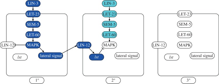
Caenorhabditis elegans vulval development is a paradigm system for understanding cell differentiation in the process of organogenesis. Through temporal and spatial controls, the fate pattern of six cells is determined by the competition of the LET-23 and the Notch signalling pathways. Modelling cell fate determination in vulval development using state-based models, coupled with formal analysis techniques, has been established as a powerful approach in predicting the outcome of combinations of mutations. However, computing the outcomes of complex and highly concurrent models can become prohibitive. Here, we show how logic programs derived from state machines describing the differentiation of C. elegans vulval precursor cells can increase the speed of prediction by four orders of magnitude relative to previous approaches. Moreover, this increase in speed allows us to infer, or 'retrodict', compatible genomes from cell fate patterns. We exploit this technique to predict highly variable cell fate patterns resulting from dig-1 reduced-function mutations and let-23 mosaics. In addition to the new insights offered, we propose our technique as a platform for aiding the design and analysis of experimental data.

Keywords: C. elegans; development; executable modelling; logic programming; organogenesis.


Figures

Figure 1.
Figure 1.
Competition between the inductive and lateral signals determines cell fate. Blue cells are 1°, red 2° and yellow 3°. Strong inductive signal from the anchor cell causes the neighbouring VPC (P6.p) to activate the LET-23/LET-60/MAPK cascade, which leads to the cell generating the lateral signal and the cell adopting the 1° fate. Adjacent cells (P5.p and P7.p) receive the lateral signal (red arrow), which inhibits the transduction of the weaker inductive signal and leads to the cell adopting the 2° cell fate. Other cells (P3.p, P4.p and P8.p) which do not receive any signals adopt the 3° fate. LIN-15 prevents secretion of LIN-3 by the hypoderm (green inhibitory arrow), and a loss of function mutation of LIN-15 causes the cells at P3.p, P4.p and P8.p to assume a non-3° fate.
Figure 2.
Figure 2.
Specific pathways of cell fate determination. Active elements are indicated in dark blue, partially active elements in cyan, and inactive elements in white. Activating relationships are indicated by an arrow, inhibitory by a blocked line. Activation of LET-23 by LIN-3 leads to downstream activation of MAPK, which generates the lateral signal (via LIN-1), and inhibits LIN-12 (via SUR-2). The lateral signal activates LIN-12 in the adjacent cells, which inhibits the LET-23/SEM-5/LET-60/MAPK cascade through multiple lateral signal target gene products (collectively termed lst genes). In 3° cells, neither pathway is activated. LIN-15 loss-of-function mutations cause LIN-3 to be secreted by the hypoderm, strongly activating LET-23 in all six VPCs.
Figure 3.
Figure 3.
Flowchart shows how LET-23 activity varies. The activity of LET-23 is dependent on a combination of the state of its gene, its location (determined by the state of lin-2), and the level of LIN-3 the cell is responding to. If let-23 is lost, or the location of LET-23 is in the apical membrane, then the activity of LET-23 is always set to none. When LET-23 is located mostly or entirely in the basolateral membrane, its activity matches the level of LIN-3 experienced by the cell; when its location is apical, its activity is always none.
Figure 4.
Figure 4.
Changes induced by the dig-1 mutation. In the wild-type, the anchor cell is always ventral and located next to P6.p. In dig-1 loss-of-function mutants where the gonad is ventral, the anchor cell can be located next to P3.p to P6.p (indicated by dashed cell edges) but otherwise behaves as wild-type. In dig-1 mutants where the gonad is dorsal, the cells each receive a variable quantity of inductive signal, regardless of anchor cell position.
Figure 5.
Figure 5.
Effects of lateral summation. Active elements are indicated in blue, weakly active elements in cyan and inactive elements in white. Activating relationships are indicated by an arrow, inhibitory by a blocked line. A cell can experience a high lateral signal if it experiences two separate medium strength lateral signals. This can arise from a cell which produces no lateral signal neighbouring two cells which generate a medium lateral signal, or if a cell generates its own medium strength lateral signal and receives a medium strength signal from its neighbour.

References

    1. Fisher J, Henzinger TA. 2007. Executable cell biology. Nat. Biotechnol. 25, 1239–1249. (10.1038/nbt1356) - DOI - PubMed
    1. Sadot A, Fisher J, Barak D, Admanit Y, Stern MJ, Hubbard EJA, Harel D. 2008. Toward verified biological models. IEEE/ACM Trans. Comput. Biol. Bioinform. 5, 223–234. (10.1109/TCBB.2007.1076) - DOI - PubMed
    1. Bonzanni N, Feenstra KA, Fokkink W, Krepska E. 2009. What can formal methods bring to systems biology? In FM 2009: formal methods, pp. 16–22. Berlin, Germany: Springer.
    1. Setty Y, Cohen IR, Dor Y, Harel D. 2008. Four-dimensional realistic modeling of pancreatic organogenesis. Proc. Natl Acad. Sci. USA 105, 20 374–20 379. (10.1073/pnas.0808725105) - DOI - PMC - PubMed
    1. Efroni S, Harel D, Cohen IR. 2003. Toward rigorous comprehension of biological complexity: modeling, execution, and visualization of thymic T-cell maturation. Genome Res. 13, 2485–2497. (10.1101/gr.1215303) - DOI - PMC - PubMed

LinkOut - more resources