. 2014 Aug 8;289(32):22524-35.
doi: 10.1074/jbc.M114.582015. Epub 2014 Jun 25.

Motor-mediated cortical versus astral microtubule organization in lipid-monolayered droplets

Affiliations

Motor-mediated cortical versus astral microtubule organization in lipid-monolayered droplets

Hella Baumann et al. J Biol Chem. .

Abstract

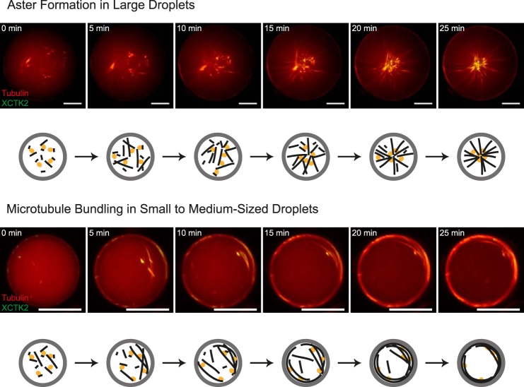
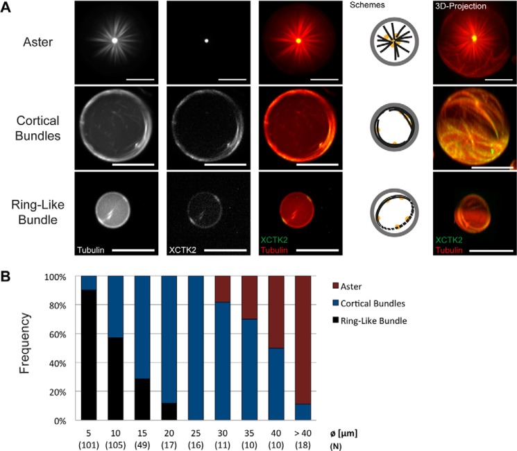
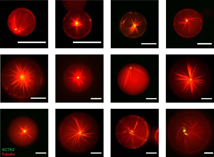
The correct spatial organization of microtubules is of crucial importance for determining the internal architecture of eukaryotic cells. Microtubules are arranged in space by a multitude of biochemical activities and by spatial constraints imposed by the cell boundary. The principles underlying the establishment of distinct intracellular architectures are only poorly understood. Here, we studied the effect of spatial confinement on the self-organization of purified motors and microtubules that are encapsulated in lipid-monolayered droplets in oil, varying in diameter from 5-100 μm, which covers the size range of typical cell bodies. We found that droplet size alone had a major organizing influence. The presence of a microtubule-crosslinking motor protein decreased the number of accessible types of microtubule organizations. Depending on the degree of spatial confinement, the presence of the motor caused either the formation of a cortical array of bent microtubule bundles or the generation of single microtubule asters in the droplets. These are two of the most prominent forms of microtubule arrangements in plant and metazoan cells. Our results provide insights into the combined organizing influence of spatial constraints and cross-linking motor activities determining distinct microtubule architectures in a minimal biomimetic system. In the future, this simple lipid-monolayered droplet system characterized here can be expanded readily to include further biochemical activities or used as the starting point for the investigation of motor-mediated microtubule organization inside liposomes surrounded by a deformable lipid bilayer.

Keywords: Confocal Microscopy; Cytoskeleton; Microtubule; Molecular Motor; Protein Self-Assembly; Reaction Compartmentalization; Self-organization.


Figures

FIGURE 1.
FIGURE 1.
Effect of lipid composition on microtubule polymerization. Lipid-monolayered droplets (in mineral oil) containing 40 μm Atto633-labeled tubulin, and 3 mm GTP in droplet buffer (see “Experimental Procedures”) with or without glycerol. Images of the equatorial plane of the droplets were recorded by spinning disk confocal microscopy 20 min after polymerization was triggered by a temperature shift to 32 °C. A, in DOPC-monolayered droplets, tubulin forms aggregates both in the absence (top panel) and presence of 10% glycerol (bottom panel). B and C, in DOPC/DOPG-monolayered droplets (B) and in DOPC/DOPE-monolayered droplets (C), tubulin neither aggregates nor polymerizes (top panels). However it polymerizes to microtubules in the presence of 10% glycerol (bottom panels). D, in DOPC/DOPE/DOPG-monolayered droplets, microtubules polymerize in the presence of 10% glycerol. Scale bars = 20 μm.
FIGURE 2.
FIGURE 2.
Categories of confined microtubule arrangements. Categorization of different microtubule arrangements inside lipid-monolayered droplets. Three-dimensional projections (top panel, top row) and images of the equatorial plane (top panel, bottom row) of Atto633-microtubules inside DOPC/DOPE/DOPG-monolayered droplets as imaged with spinning disk confocal microscopy 20 min after the polymerization reaction was started. The arrangements at end state were classified into a network of evenly distributed microtubules (A), a cortical arrangement (B), an opening cloud (C), a ring-like bundle (D), or a spoke-like arrangement (E). Schemes (bottom panel) show an interpretation for each arrangement type of microtubules (black) inside the lipid-monolayered droplets (gray). The tubulin concentration was 40 μm.
FIGURE 3.
FIGURE 3.
Time course of confined microtubule arrangements: characteristic examples for the different arrangement categories. De novo polymerization and formation of different arrangements of microtubules inside DOPC/DOPE/DOPG-monolayered droplets. Representative time series of spinning disk confocal microscopy images of confined microtubules polymerizing from 40 μm Atto633-tubulin in standard droplet buffer in response to a temperature shift to 32 °C. Examples for the different categories of arrangements are shown: network of evenly distributed microtubules (A), cortical arrangement (B), ring-like bundle (C), spoke-like arrangement (D), and opening cloud of microtubules (E). Schematics of the arrangements are shown below the experimental data. Scale bars = 20 μm.
FIGURE 4.
FIGURE 4.
Droplet size distribution and frequency of microtubule arrangements over droplet size. A, histogram of the size distribution of observed DOPC/DOPE/DOPG-monolayered droplets. The bar graph displays the relative frequencies of the categories of formed microtubule arrangements after 20 min in droplets of different size, containing 40 (B), 30 (C), or 50 μm (D) tubulin in standard droplet buffer. Droplets were assigned to the diameter bins (Ø) as indicated. Numbers in parentheses indicate the total count (N) of droplets for each size category.
FIGURE 5.
FIGURE 5.
Time course of confined motor-driven microtubule self-organization. Shown is motor-driven microtubule self-organization inside DOPC/DOPE/DOPG-monolayered droplets of different sizes. Also shown is a time series of characteristic images of motor-driven microtubule organization inside a large (85-μm diameter, top panel) and a medium-sized (30-μm diameter, bottom panel) lipid-monolayered droplet containing 40 μm Atto633-labeled tubulin (red) and 200 nm mCherry-kinesin-14 (green) in standard droplet buffer, as acquired with spinning disk confocal microscopy at 32 °C. Schematics of the observed microtubule (black) and motor (orange) organizations inside droplets (gray) are shown below the experimental data. Scale bars = 20 μm.
FIGURE 6.
FIGURE 6.
Droplet size dependence of confined motor-driven microtubule self-organization. A, different categories of end states. Spinning disk confocal microscopy images of DOPC/DOPE/DOPG-monolayered droplets containing 40 μm Atto633-labeled tubulin (red in merge) and 200 nm mCherry-kinesin-14 (green in merge) in standard droplet buffer taken 25 min after reaction start are shown. Single channel and merged channel images of the equatorial droplet plane are shown as indicated. The schematics display the arrangements of microtubules (black) and the localization of the motor protein (orange) inside lipid-monolayered droplets (gray). The images on the right show three-dimensional projections. Scale bars = 20 μm. B, bar graph displaying the relative frequencies of the motor-organized microtubule organizations for different droplet diameters (Ø). Numbers in parentheses indicate the total count (N) of droplets analyzed for each size category.
FIGURE 7.
FIGURE 7.
Confined motor/microtubule self-organization into asters. Merged spinning disk confocal microscopy images of large DOPC/DOPE/DOPG-monolayered droplets containing 40 μm Atto633-labelled tubulin (red) and 200 nm mCherry-XCTK2 (green) in standard droplet buffer taken 25 min after reaction start are shown. Scale bars = 20 μm.

References

    1. Deinum E. E., Mulder B. M. (2013) Modelling the role of microtubules in plant cell morphology. Curr. Opin. Plant Biol. 16, 688–692 - PubMed
    1. Dogterom M., Surrey T. (2013) Microtubule organization in vitro. Curr. Opin. Cell Biol. 25, 23–29 - PubMed
    1. Helmke K. J., Heald R., Wilbur J. D. (2013) Interplay between spindle architecture and function. Int. Rev. Cell Mol. Biol. 306, 83–125 - PubMed
    1. Hentrich C., Surrey T. (2010) Microtubule organization by the antagonistic mitotic motors kinesin-5 and kinesin-14. J. Cell Biol. 189, 465–480 - PMC - PubMed
    1. Nédélec F. J., Surrey T., Maggs A. C., Leibler S. (1997) Self-organization of microtubules and motors. Nature 389, 305–308 - PubMed

Publication types

LinkOut - more resources