. 2014 Oct;12(10):1460-9.
doi: 10.1158/1541-7786.MCR-14-0038. Epub 2014 Jun 25.

Nonamplified FGFR1 is a growth driver in malignant pleural mesothelioma

Affiliations

Nonamplified FGFR1 is a growth driver in malignant pleural mesothelioma

Lindsay A Marek et al. Mol Cancer Res. 2014 Oct.

Abstract

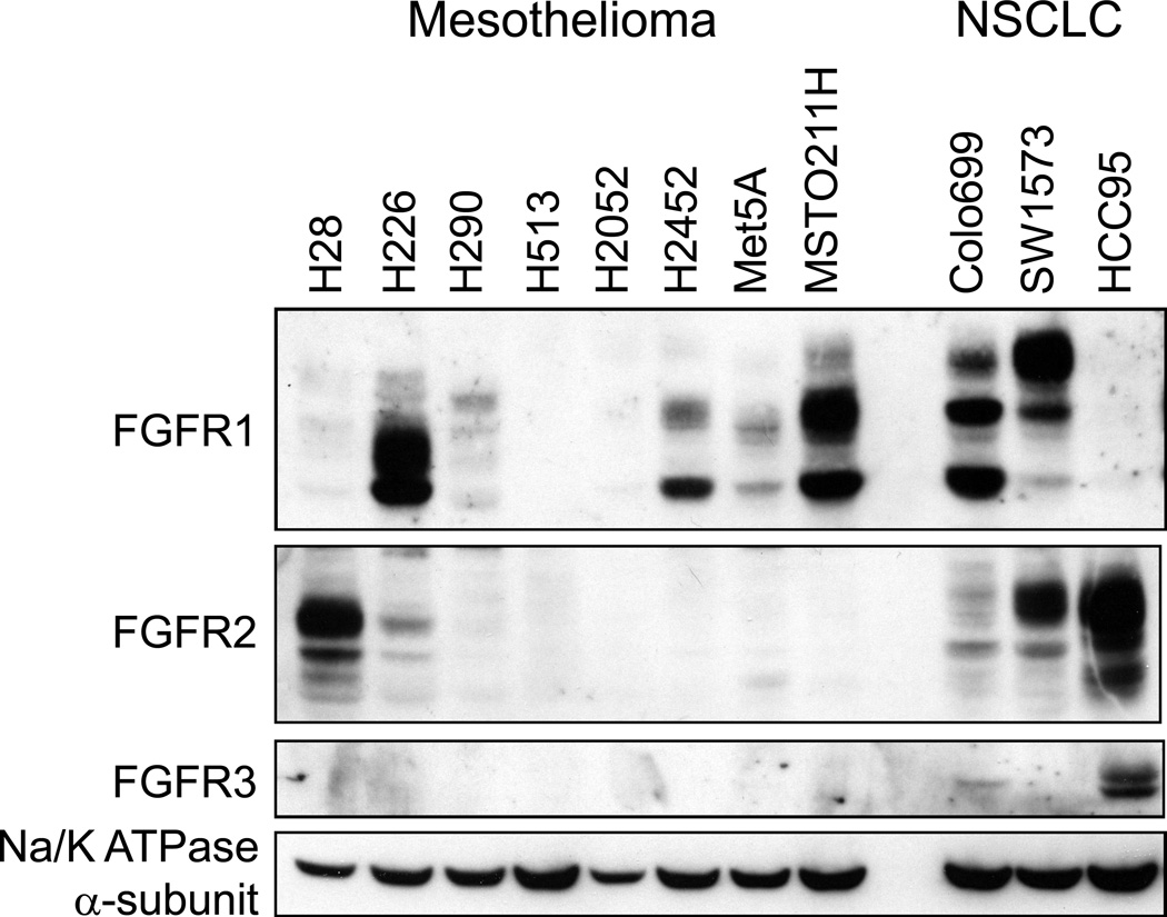
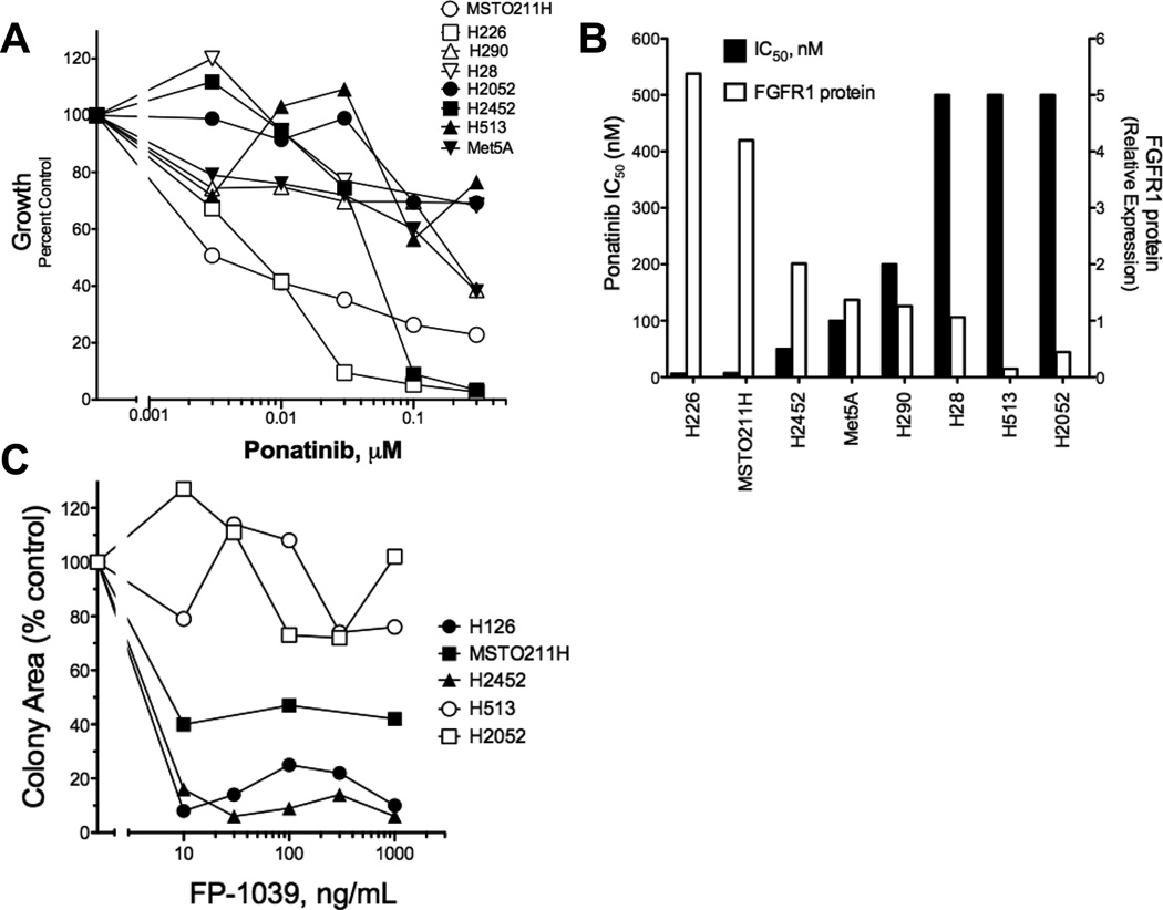
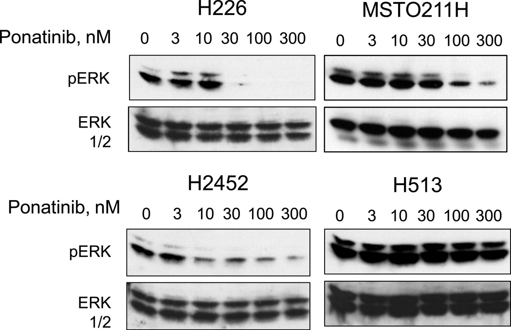
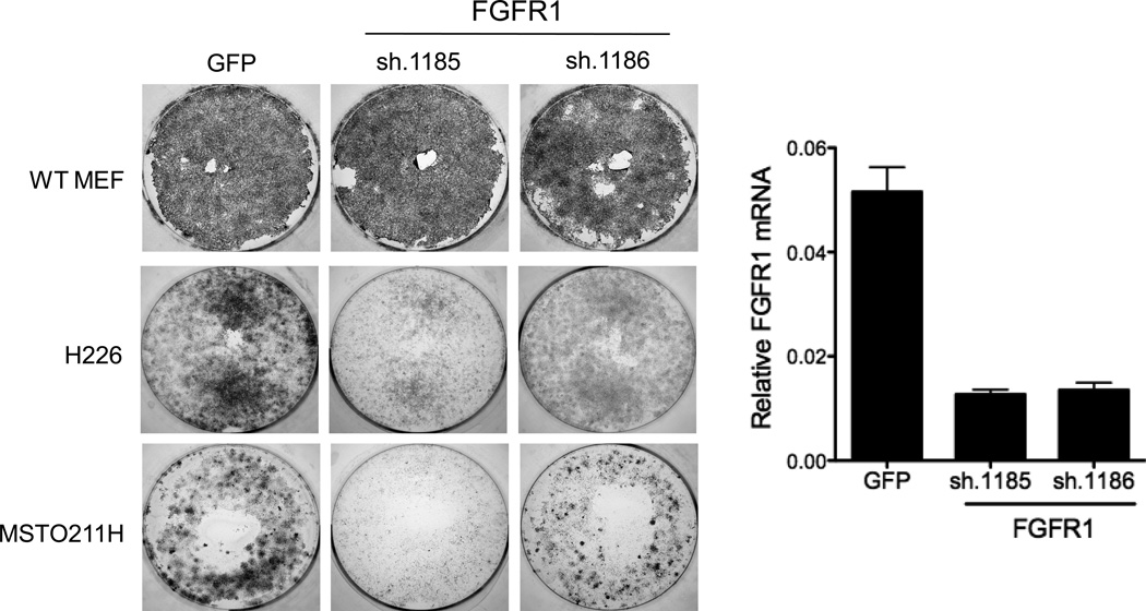
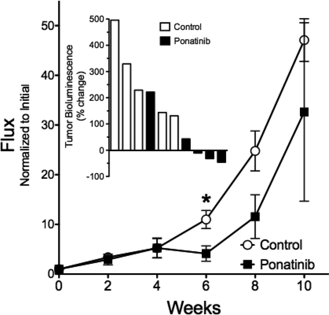
Malignant pleural mesothelioma (MPM) is associated with asbestos exposure and is a cancer that has not been significantly affected by small molecule-based targeted therapeutics. Previously, we demonstrated the existence of functional subsets of lung cancer and head and neck squamous cell carcinoma (HNSCC) cell lines in which fibroblast growth factor receptor (FGFR) autocrine signaling functions as a nonmutated growth pathway. In a panel of pleural mesothelioma cell lines, FGFR1 and FGF2 were coexpressed in three of seven cell lines and were significantly associated with sensitivity to the FGFR-active tyrosine kinase inhibitor (TKI), ponatinib, both in vitro and in vivo using orthotopically propagated xenografts. Furthermore, RNAi-mediated silencing confirmed the requirement for FGFR1 in specific mesothelioma cells and sensitivity to the FGF ligand trap, FP-1039, validated the requirement for autocrine FGFs. None of the FGFR1-dependent mesothelioma cells exhibited increased FGFR1 gene copy number, based on a FISH assay, indicating that increased FGFR1 transcript and protein expression were not mediated by gene amplification. Elevated FGFR1 mRNA was detected in a subset of primary MPM clinical specimens and like MPM cells; none harbored increased FGFR1 gene copy number. These results indicate that autocrine signaling through FGFR1 represents a targetable therapeutic pathway in MPM and that biomarkers distinct from increased FGFR1 gene copy number such as FGFR1 mRNA would be required to identify patients with MPM bearing tumors driven by FGFR1 activity.

Implications: FGFR1 is a viable therapeutic target in a subset of MPMs, but FGFR TKI-responsive tumors will need to be selected by a biomarker distinct from increased FGFR1 gene copy number, possibly FGFR1 mRNA or protein levels.


Conflict of interest statement

Disclosure of Potential Conflicts of Interest: L. Heasley is the recipient of a research contract with ARIAD Pharmaceuticals, Inc to complete pre-clinical studies with ponatinib.

Figures

Figure 1
Figure 1. FGFR protein expression in mesothelioma cell lines
Cell extracts were prepared from the indicated cell lines and aliquots were submitted to SDS-PAGE. Following electrophoretic transfer, the filters were probed for antibodies to FGFR1–3 and the α-subunit of NaK-ATPase as a loading control. Colo699 and SW1734 cells were employed as positive controls for FGFR1 and HCC95 cells are a positive control for expression of FGFR2 and FGFR3.
Figure 2
Figure 2. Inhibition of in vitro growth of mesothelioma cell lines by the FGFR inhibitor, ponatinib
A, Anchorage-independent growth (H2052, MSTO211H, H513, H226, Met5A, H2452) or cell proliferation assays over 7 days (H28, H290) were performed in the presence of the indicated concentrations of ponatinib as described in the Materials and Methods. B, The IC50 values were calculated with Prism software (GraphPad, San Diego, CA) and plotted versus FGFR1 protein expression measured by densitometry of immunoblots. C, Anchorage-independent growth of the indicated cell lines was measured in the presence of increasing concentrations of the FGF trap, FP-1039 and the data are presented as growth as a percent of control growth measured in the absence of FP-1039.
Figure 3
Figure 3. Inhibition of basal ERK activity in H226, MSTO211H and H2452 cells by ponatinib
The indicated cell lines were incubated in serum-free medium for 2 hours in the presence of increasing concentrations of ponatinib. Cell extracts were prepared and immunoblotted for phospho-ERK and as described in the Materials and Methods. The filters were stripped and reprobed for total ERK to verify equal protein loading.
Figure 4
Figure 4. Inhibition of clonogenic growth following RNAi-mediated silencing of FGFR1
Lentiviral-encoded shRNAs targeting GFP as a non-silencing control or two independent shRNAs against FGFR1 were packaged and transduced into H226 and MSTO211H cells and then selected for puromycin resistance. Mouse embryo fibroblasts (MEFs) were similarly transduced as a test for the relative titer of the different lentiviral preparations. Following 8 days, the cultures were fixed, stained with crystal violet and photographed. Total RNA was prepared from replicate cultures and following reverse-transcription to cDNA, was submitted to a quantitative PCR assay for FGFR1 and GAPDH mRNAs as described in the Materials and Methods.
Figure 5
Figure 5. Effect of ponatinib on orthotopic growth of H226 cells in immune-deficient mice
Luciferase-expressing H226 cells (106) were implanted into the pleural space of immune-deficient mice as described in the Materials and Methods. Six weeks later (t = 0 on graph), the tumor-bearing mice were randomized into groups of 5 mice and treated by oral gavage with ponatinib (20 mg/kg) or diluent daily. The mice were submitted to bioluminescence imaging (BLI) every two weeks to determine tumor burden and the data are presented as the photon flux normalized to the initial tumor measurement with the means and SEM shown; * indicates p<0.05 by student’s two-tailed t-test. The inset graph shows the percent change in tumor bioluminescence measured at six weeks of treatment relative to the bioluminescence observed at the initiation of treatment for the individual control and ponatinib-treated mice where 3 of 5 ponatinib-treated mice exhibited tumor shrinkage.

References

    1. Raja S, Murthy SC, Mason DP. Malignant pleural mesothelioma. Curr Oncol Rep. 2011;13:259–264. - PubMed
    1. van der Bij S, Koffijberg H, Burgers JA, Baas P, van de Vijver MJ, de Mol BA, et al. Prognosis and prognostic factors of patients with mesothelioma: a population-based study. Br J Cancer. 2012;107:161–164. - PMC - PubMed
    1. Becklake MR, Bagatin E, Neder JA. Asbestos-related diseases of the lungs and pleura: uses, trends and management over the last century. Int J Tuberc Lung Dis. 2007;11:356–369. - PubMed
    1. Ceresoli GL, Locati LD, Ferreri AJ, Cozzarini C, Passoni P, Melloni G, et al. Therapeutic outcome according to histologic subtype in 121 patients with malignant pleural mesothelioma. Lung Cancer. 2001;34:279–287. - PubMed
    1. Yates DH, Corrin B, Stidolph PN, Browne K. Malignant mesothelioma in south east England: clinicopathological experience of 272 cases. Thorax. 1997;52:507–512. - PMC - PubMed

Publication types

MeSH terms