. 2014 Jul;42(13):8755-66.
doi: 10.1093/nar/gku554. Epub 2014 Jun 25.

Transition-state destabilization reveals how human DNA polymerase β proceeds across the chemically unstable lesion N7-methylguanine

Affiliations

Transition-state destabilization reveals how human DNA polymerase β proceeds across the chemically unstable lesion N7-methylguanine

Myong-Chul Koag et al. Nucleic Acids Res. 2014 Jul.

Abstract

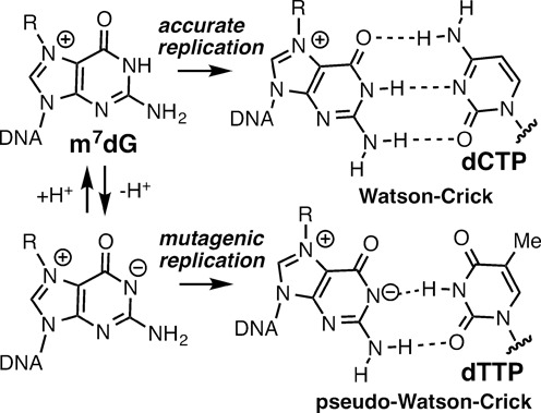
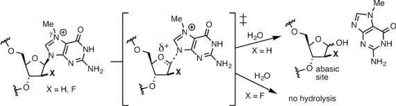
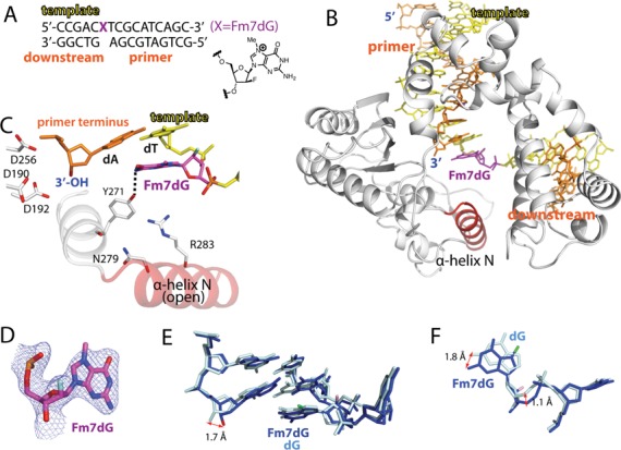
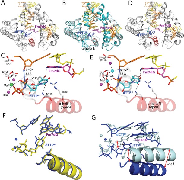
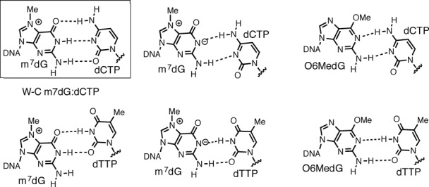
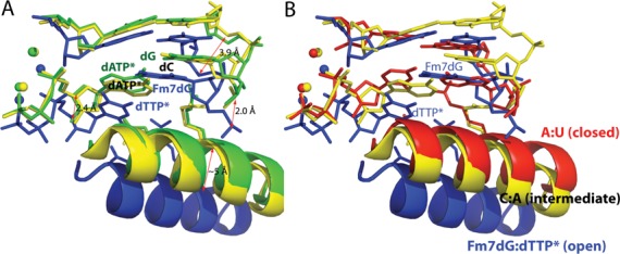
N7-Methyl-2'-deoxyguanosine (m7dG) is the predominant lesion formed by methylating agents. A systematic investigation on the effect of m7dG on DNA replication has been difficult due to the chemical instability of m7dG. To gain insights into the m7dG effect, we employed a 2'-fluorine-mediated transition-state destabilzation strategy. Specifically, we determined kinetic parameters for dCTP insertion opposite a chemically stable m7dG analogue, 2'-fluoro-m7dG (Fm7dG), by human DNA polymerase β (polβ) and solved three X-ray structures of polβ in complex with the templating Fm7dG paired with incoming dCTP or dTTP analogues. The kinetic studies reveal that the templating Fm7dG slows polβ catalysis ∼ 300-fold, suggesting that m7dG in genomic DNA may impede replication by some DNA polymerases. The structural analysis reveals that Fm7dG forms a canonical Watson-Crick base pair with dCTP, but metal ion coordination is suboptimal for catalysis in the polβ-Fm7dG:dCTP complex, which partially explains the slow insertion of dCTP opposite Fm7dG by polβ. In addition, the polβ-Fm7dG:dTTP structure shows open protein conformations and staggered base pair conformations, indicating that N7-methylation of dG does not promote a promutagenic replication. Overall, the first systematic studies on the effect of m7dG on DNA replication reveal that polβ catalysis across m7dG is slow, yet highly accurate.


Figures

Figure 1.
Figure 1.
Structures of Watson–Crick m7dG:dCTP and pseudo-Watson–Crick m7dG:dTTP base pairs.
Figure 2.
Figure 2.
Inhibition of depurination of m7dG via 2′-F-mediated transition-state destabilization
Figure 3.
Figure 3.
Single-nucleotide gapped binary structure of polβ in complex with DNA containing templating Fm7dG (PDB ID: 4O5C). (A) The DNA sequence used for crystallography and chemical structure of Fm7dG are shown. The downstream primer contains 5′-phosphate. (B) Overall crystal structure of the Fm7dG gapped binary complex. Polβ is shown in white. The α-helix N bearing the minor groove recognition amino acid residues Asn279 and Arg283 is shown in red. The template strand is shown in yellow and the upstream and the downstream primers are shown in orange. The templating Fm7dG is indicated. (C) Active-site structure. The α-helix N is in an open conformation. Y271 is H-bonded to N2 of Fm7dG (D) A 2Fo-Fc electron density map is contoured at 1σ around Fm7dG. Templating Fm7dG adopts the 2′-endo sugar pucker and the anti-base conformation. (E) Comparison of the Fm7dG gapped structure (blue) with published dG gapped structure (pale cyan, PDB ID: 1BPX). Distance between the 3′-OHs of the primer terminus is indicated. (F) Comparison of templating Fm7dG (blue) with templating dG (pale cyan) in (E).
Figure 4.
Figure 4.
Ternary structure of polβ in complex with templating Fm7dG paired with an incoming nonhydrolyzable dCTP analog (PDB ID: 4O5K). (A) Overall structure of the Fm7dG:dCTP* complex. The protein adopts the closed conformation and the nascent base pair adopts a coplanar conformation. (B) Close-up view of the active-site structure. The α-helix N is in a closed conformation. Key interactions are indicated as dotted lines. Active-site Mg2+ ions are shown as green spheres, and water molecules are shown as magenta spheres. Note that 3′-OH of the primer terminus is not coordinated to the catalytic metal ion. The distance between 3′-OH of the primer terminus and Pα of the incoming nucleotide is indicated. (C) Base-pairing mode of Fm7dG:dCTP*. A 2FoFc electron density map is contoured at 1σ around Fm7dG and dCTP*. The H-bonding distances, the C1(dCTP)′ − C1(Fm7dG)′ distance and λ angles are very similar to those observed in a polβ structure with the correct insertion (PDB ID: 2FMS). (D) Overlay of the Fm7dG:dTTP*–Mg2+ ternary structure with published polβ ternary structure (PDB ID: 2FMS) with nascent dA:dUTP* base pair (RMSD = 0.400 Å) (E) Overlay of the active site of the Fm7dG:dTTP*–Mg2+ structure (blue) and published dA:dUTP* ternary structure (pale cyan). (F) Top view of the Fm7dG:dTTP* base pair (blue) and the dA:dUTP* base pair (pale cyan) in (E). (G) Overlay of DNA in the Fm7dG:dTTP*–Mg2+ structure (blue) and published dA:dUTP* ternary structure (pale cyan).
Figure 5.
Figure 5.
Ternary structure of polβ with templating Fm7dG paired with an incoming nonhydrolyzable dTTP analog (PDB ID: 4O5E and 4P2H). (A) Overall structure of the Fm7dG:dTTP*–Mg2+ complex. The protein adopts an open conformation, and the nascent base pair adpots a staggered conformation. (B) Overlay of the Fm7dG:dTTP*–Mg2+ structure (white) with the Fm7dG gapped binary structure (cyan) (RMSD = 0.316 Å). (C) Close-up view of the active-site structure of the Fm7dG:dTTP*–Mg2+ complex. Fm7dG does not H-bond with the incoming dTTP*. Only the nucleotide-binding metal ion is observed. (D) Overall structure of the Fm7dG:dTTP*–Mn2+ complex. (E) Active-site structure of the Fm7dG:dTTP*–Mn2+ complex. Both the nucleotide-binding and the catalytic metal ions are present. (F) Comparison of the active site structure of the Fm7dG:dTTP*–Mn2+ complex (blue) with that of the Fm7dG:dTTP*–Mg2+ complex (yellow). (G) Comparison of the active site structure of the Fm7dG:dTTP*–Mn2+ complex (blue) with that of the Fm7dG:dCTP*–Mg2+ complex (pale cyan).
Figure 6.
Figure 6.
Potential base pairings of a modified dG with an incoming dCTP/dTTP nucleotide. (A) Base pairings of m7dG with dCTP/dTTP (B) Base pairings of O6-methyl-dG (O6MedG) with dCTP/dTTP. Base pairs observed in the Fm7dG structures and published O6-methyl-dG structures are shown in a box. A polβ structure with the wobble base pair conformation has not been observed.
Figure 7.
Figure 7.
Comparison of the Fm7dG:dTTP*–Mn2+ structure with published mismatched structures. Overlay of the Fm7dG:dTTP*–Mn2+ structure (blue) with published: (A) dC:dATP–Mn2+ structure (yellow, PDB ID: 3C2L) and dG:dATP*–Mn2+ structure (green, PDB ID: 3C2M), (B) dC:dATP*–Mn2+ structure (yellow, PDB ID: 3C2L) and dA:dUTP*–Mg2+ structure (red, PDB ID: 2FMS).

References

    1. Lindahl T. Instability and decay of the primary structure of DNA. Nature. 1993;362:709–715. - PubMed
    1. Sedgwick B. Repairing DNA-methylation damage. Nat. Rev. Mol. Cell Biol. 2004;5:148–157. - PubMed
    1. Reiner B., Zamenhof S. Studies on the chemically reactive groups of deoxynucleic acids. J. Biol. Chem. 1957;228:475–486. - PubMed
    1. Hayashibara K.C., Verdine G.L. Template-directed interference footprinting of cytosine contacts in a protein-DNA complex: potent interference by 5-aza-2’-deoxycytidine. Biochemistry. 1992;31:11265–11273. - PubMed
    1. Ezaz-Nikpay K., Verdine G.L. Aberrantly methylated DNA: site-specific introduction of N-7-methyl-2’-deoxyguanosine into the Dickerson/Drew dodecamer. J. Am. Chem. Soc. 1992;114:6562–6563.

Publication types