. 2014 Jun 25;34(26):8749-60.
doi: 10.1523/JNEUROSCI.0141-14.2014.

RAGE inhibition in microglia prevents ischemia-dependent synaptic dysfunction in an amyloid-enriched environment

Affiliations

RAGE inhibition in microglia prevents ischemia-dependent synaptic dysfunction in an amyloid-enriched environment

Nicola Origlia et al. J Neurosci. .

Abstract

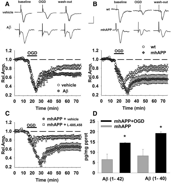
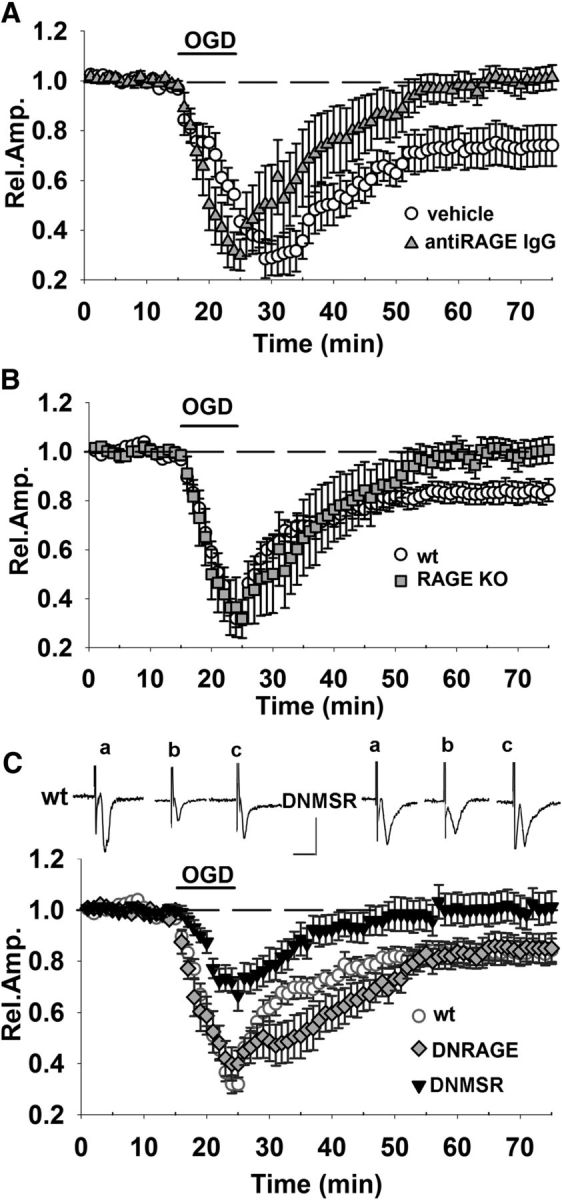
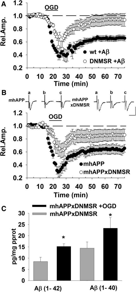
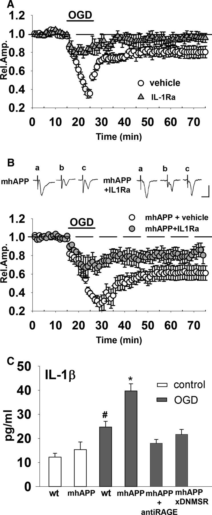
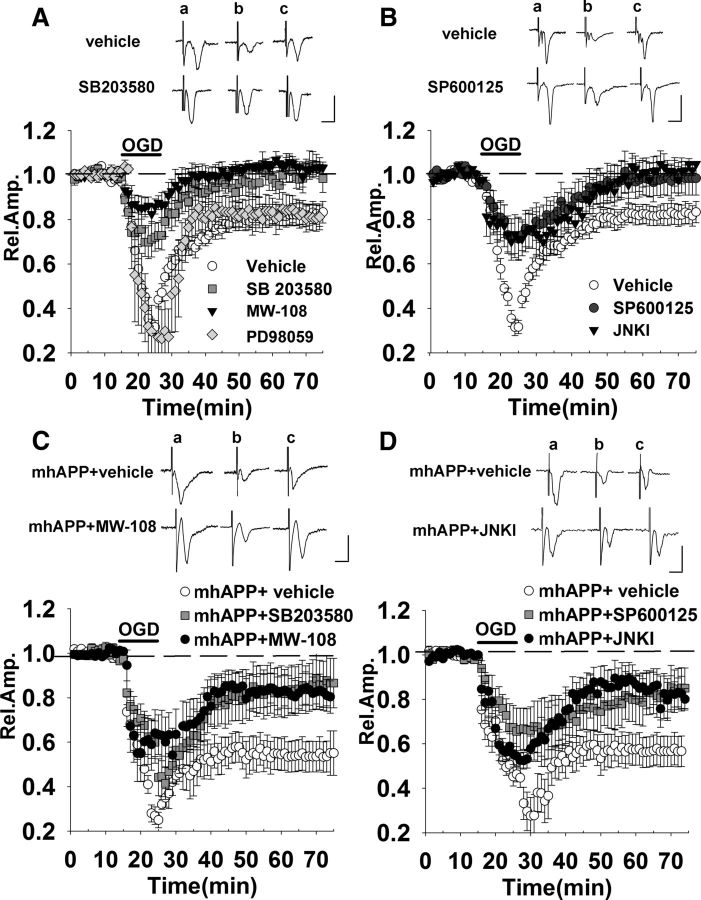
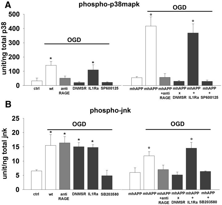
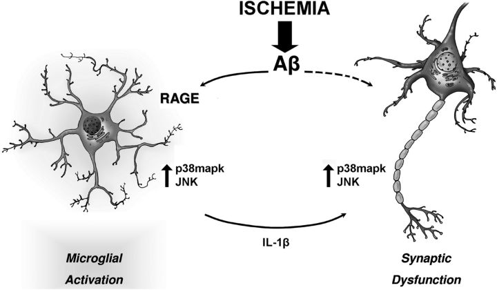
Ischemia is known to increase the deleterious effect of β-amyloid (Aβ), contributing to early cognitive impairment in Alzheimer's disease. Here, we investigated whether transient ischemia may function as a trigger for Aβ-dependent synaptic impairment in the entorhinal cortex (EC), acting through specific cellular signaling. We found that synaptic depression induced by oxygen glucose deprivation (OGD) was enhanced in EC slices either in presence of synthetic oligomeric Aβ or in slices from mutant human amyloid precursor protein transgenic mice (mhAPP J20). OGD-induced synaptic depression was ameliorated by functional suppression of RAGE. In particular, overexpression of the dominant-negative form of RAGE targeted to microglia (DNMSR) protects against OGD-induced synaptic impairment in an amyloid-enriched environment, reducing the activation of stress-related kinases (p38MAPK and JNK) and the release of IL-1β. Our results demonstrate a prominent role for the RAGE-dependent neuroinflammatory pathway in the synaptic failure induced by Aβ and triggered by transient ischemia.

Keywords: entorhinal cortex; interleukin1-beta; jnk; neuroinflammation; p38mapk; synaptic transmission.


Figures

Figure 1.
Figure 1.
OGD-dependent synaptic depression is enhanced in the presence of Aβ. A, OGD for 10 min (corresponding to dark bar) induced a decrease of FP amplitude in control vehicle-treated slices (open circles), reaching a stable level of depression after 50 min. OGD-induced depression was enhanced in slices treated with Aβ(1-42) at 1 μm (gray circles) for 10 min (corresponding to dark bar). Insets show typical traces of FP recordings before during and after OGD exposure in vehicle- and Aβ-treated slices. B, Synaptic depression after 10 min of OGD (dark bar) was significantly increased in mhAPP slices (gray diamonds) with respect to WT (open circles). Insets show typical traces of FP recordings before, during, and after OGD in WT or mhAPP slices. C, OGD (dark bar, 10 min)-induced depression was significantly reduced in mhAPP slices continuously perfused with 0.5 μm l-685,458 (white squares) with respect to vehicle-treated mhAPP slices. D, Bars represent the average Aβ(1-40) (right) and Aβ(1-42) (left) levels measured using an ELISA assay. Both Aβ(1-40) and Aβ(1-42) were significantly elevated (#p < 0.05) in mhAPP slices exposed to OGD with respect to control nonexposed mhAPP slices. Scale bars: A, B, horizontal, 5 ms; vertical, 0.5 mV. Error bars indicate SEM.
Figure 2.
Figure 2.
Microglial RAGE contributes to OGD-induced synaptic depression. Blockade of RAGE by either knocking out the RAGE gene or with neutralizing anti-RAGE IgG was able to prevent ischemia-dependent long-lasting depression. FP amplitude in anti-RAGE IgG (A, gray triangles) and RAGE-KO (B, gray squares) slices was completely rescued 50 min after OGD (10 min corresponding to dark bar) and was significantly different from FP amplitude in OGD-exposed WT (white circles). C, RAGE-deficient signaling in neurons (DN-RAGE, gray diamonds) was not sufficient to prevent OGD-induced synaptic depression. In contrast, DNMSR slices (c, black triangles), characterized by RAGE signaling deficiency in microglia, displayed a significant reduction of synaptic depression during OGD (10 min, dark bar) compared with WT (white circles) and a full recovery of FP amplitude was achieved 50 min after. Insets show typical traces of FP recordings before (a), during (b), and after (c) OGD in WT or DNMSR slices. Scale bars, 10 ms (horizontal) and 1 mV (vertical). WT slices recorded in interleaved experiments were pooled together and reported as one group in B and C. In A, vehicle control solution contained rabbit nonimmune IgG.
Figure 3.
Figure 3.
Microglial RAGE inhibition protects against OGD-induced synaptic dysfunction in an Aβ-enriched environment. A, Effect of OGD (dark bar, 10 min) coupled to exogenous supply of Aβ(1-42) (1 μm perfused for 10 min) in slices from DNMSR mice (gray diamonds) was significantly reduced with respect to Aβ-treated WT slices exposed to OGD (black circles). B, A deficiency of RAGE in microglia was able to counteract the effect of OGD in the presence of mutant APP overexpression. The synaptic depression was significantly lower in slices from double-transgenic mice (DNMSR × mhAPP, white circles) with respect to single mhAPP transgenic slices (black circles). Insets show typical traces of FP recordings before (a), during (b), and after (c) OGD in mhAPP or mhAPPxDNMSR slices. Scale bars, 10 ms (horizontal) and 1 mV (vertical). C, Bars represent the average Aβ(1-40) (right) and Aβ(1-42) (left) levels measured using an ELISA assay. Both Aβ(1-40) and Aβ(1-42) were significantly elevated (*p < 0.05) in mhAPPxDNMSR slices exposed to OGD with respect to control nonexposed mhAPPxDNMSR slices.
Figure 4.
Figure 4.
IL1β is increased during OGD and its blockade by IL1Ra reduces synaptic dysfunction in mhAPP slices. A, When slices were treated with 20 ng/ml IL-1Ra (gray triangles), synaptic depression during OGD (10 min, dark bar) was significantly decreased with respect to OGD-exposed vehicle-treated slices. In the presence of IL-1Ra (gray triangles), OGD failed to induce long-lasting synaptic depression of FP amplitude that returned to baseline values 50 min after transient ischemia. B, Synaptic depression caused by OGD (dark bar) was significantly reduced by IL1Ra pretreatment in mhAPP slices (gray circles) compared with vehicle-treated mhAPP slices (white circles). Insets show typical traces of FP recordings before (a), during (b), and after (c) OGD in mhAPP vehicle-treated or mhAPP IL1Ra-treated slices. Scale bars, 10 ms (horizontal) and 1 mV (vertical). C, Plot representing IL-1β levels in EC slices exposed to OGD. The basal levels of IL-1β did not differ between WT and mhAPP mice (p < 0.05), but were significantly elevated in WT slices exposed to OGD compared with nonexposed WT and mhAPP slices (#p < 0.01). Ten minutes of OGD induced a significantly higher level of IL-1β in mhAPP slices compared with nonexposed mhAPP (*p < 0.001) and this increase was significantly higher than that found in WT slices exposed to OGD (*p < 0.001). Error bars indicate SEM.
Figure 5.
Figure 5.
OGD-induced synaptic dysfunction in WT and mhAPP slices is prevented by inhibition of the stress-related kinases p38MAPK and JNK. A, Treatment of slices with either 1 μm SB203580 (gray squares) or 10 μm MW-108 (filled triangles) was able to reduce synaptic depression during OGD (dark bar) and allowed a full recovery of FPs 50 min after transient ischemia. In contrast, the MEK inhibitor PD 98059 (50 μm, gray diamonds) did not affect OGD-dependent depression. B, A similar protective effect was achieved in slices after treatment with either 20 μm SP600125 (black triangles) or JNKI (filled triangles). C, In mhAPP slices, the OGD-induced depression was significantly diminished by SB203580 (gray squares) or MW-108 (black circles) compared with vehicle-treated mhAPP slices (white circles). D, SP600125 (black triangles) and JNKI (black circles) were capable of reducing OGD-induced depression in mhAPP slices compared with vehicle-treated mhAPP slices (white circles). The insets in AD represent typical traces of FP recordings before (a), during (b), and after (c) OGD. Scale bars, 5 ms (horizontal) and 1 mV (vertical).
Figure 6.
Figure 6.
OGD increases p38MAPK/JNK phosphorylation in WT and mhAPP cortical slices. A, Plot representing averaged phospho-p38MAPK levels measured using ELISA and expressed as units per total content of p38MAPK protein. In WT slices, tissue levels of phospho-p38MAPK after 10 min of OGD were significantly higher with respect to control vehicle-treated slices (left, *p < 0.01). Selective deficiency of RAGE signaling in microglia (DNMSR) and complete inactivation of RAGE with anti-RAGE IgG was able to prevent the OGD-induced increase of phospho-p38MAPK (left, p < 0.05 vs WT OGD-exposed slices). p38MAPK activation by OGD was also reduced in presence of JNK inhibition (left, SP600125, p < 0.05 vs OGD-exposed WT slices), but not by treatment with IL-1Ra (left, *p < 0.05 vs WT control slices). A robust increase in phospho-p38MAPK was also observed in mhAPP slices after OGD (right, °p < 0.001 vs non-OGD mhAPP) and this increase was prevented in double-transgenic DNMSRxmhAPP or in anti-RAGE IgG-treated mhAPP slices (right, p > 0.05 vs non-OGD mhAPP slices). In mhAPP slices, JNK inhibition (right, SP600125) was effective in reducing p38MAPK activation, whereas phosphor-p38 was still elevated after IL-1Ra treatment (right, °p < 0.001 vs non-OGD mhAPP slices). B, Plot representing averaged phospho-JNK levels measured using ELISA and expressed as units/total content of JNK protein. Tissue levels of phospho-JNK were elevated after 10 min of OGD in WT cortical slices (left, *p < 0.05 vs WT nonexposed slices). Inhibition of RAGE (in DNMSR or anti-RAGE IgG-treated slices) or blockade of IL-1β signaling with IL-1Ra did not reduce the OGD effect (left, *p < 0.05 vs WT nonexposed slices), whereas inhibition of p38MAPK (SB203580) significantly reduced JNK activation by OGD (left, p > 0.05 vs WT nonexposed slices). A similar increase of JNK activation was observed in mhAPP slices exposed to OGD (right, °p < 0.05 vs mhAPP nonexposed slices). Blockade of RAGE by IgG or inhibition of RAGE in microglia in double-transgenic DNMSRxmhAPP mice suppressed OGD-induced increase of phospho-JNK (right, p > 0.05 vs nonexposed mhAPP slices). Basal levels of phospho-JNK were also maintained in mhAPP slices exposed to OGD that were treated with p38MAPK inhibitor (right, SB203580, p > 0.05 vs nonexposed mhAPP slices), whereas phospho-JNK was still activated in mhAPP slices treated with IL-1Ra and exposed to OGD (°p < 0.05 vs mhAPP nonexposed slices). Error bars indicate SEM.
Figure 7.
Figure 7.
Schematic drawing representing the interaction of neurons and microglia that is triggered by OGD in an amyloid-enriched environment. An ischemic insult enhances the deleterious effect of amyloid on synaptic function. Amyloid overproduction induced by OGD leads to RAGE engagement in microglia and activation of p38mapk and JNK. The kinases, in turn, control the release of proinflammatory cytokines such as IL-1β, which may affect synaptic function. This is a self-maintained loop that amplifies the effect of Aβ and shift synaptic activity toward depression.

Comment in

References

    1. Arancio O, Zhang HP, Chen X, Lin C, Trinchese F, Puzzo D, Liu S, Hegde A, Yan SF, Stern A, Luddy JS, Lue LF, Walker DG, Roher A, Buttini M, Mucke L, Li W, Schmidt AM, Kindy M, et al. RAGE potentiates Abeta-induced perturbation of neuronal function in transgenic mice. EMBO J. 2004;23:4096–4105. doi: 10.1038/sj.emboj.7600415. - DOI - PMC - PubMed
    1. Benakis C, Bonny C, Hirt L. JNK inhibition and inflammation after cerebral ischemia. Brain Behav Immun. 2010;24:800–811. doi: 10.1016/j.bbi.2009.11.001. - DOI - PubMed
    1. Braak H, Braak E. Demonstration of amyloid deposits and neurofibrillary changes in whole brain sections. Brain Pathol. 1991;1:213–216. - PubMed
    1. Chen C, Li XH, Tu Y, Sun HT, Liang HQ, Cheng SX, Zhang S. Aβ-AGE aggravates cognitive deficit in rats via RAGE pathway. Neuroscience. 2014;257:1–10. doi: 10.1016/j.neuroscience.2013.10.056. - DOI - PubMed
    1. Chen X, Walker DG, Schmidt AM, Arancio O, Lue LF, Yan SD. RAGE: a potential target for Abeta-mediated cellular perturbation in Alzheimer's disease. Curr Mol Med. 2007;7:735–742. doi: 10.2174/156652407783220741. - DOI - PubMed

Publication types

MeSH terms

Substances

LinkOut - more resources