. 2014 Jun 25;34(26):8761-71.
doi: 10.1523/JNEUROSCI.3965-13.2014.

Roles of ON cone bipolar cell subtypes in temporal coding in the mouse retina

Affiliations

Roles of ON cone bipolar cell subtypes in temporal coding in the mouse retina

Tomomi Ichinose et al. J Neurosci. .

Abstract

In the visual system, diverse image processing starts with bipolar cells, which are the second-order neurons of the retina. Thirteen subtypes of bipolar cells have been identified, which are thought to encode different features of image signaling and to initiate distinct signal-processing streams. Although morphologically identified, the functional roles of each bipolar cell subtype in visual signal encoding are not fully understood. Here, we investigated how ON cone bipolar cells of the mouse retina encode diverse temporal image signaling. We recorded bipolar cell voltage changes in response to two different input functions: sinusoidal light and step light stimuli. Temporal tuning in ON cone bipolar cells was diverse and occurred in a subtype-dependent manner. Subtypes 5s and 8 exhibited low-pass filtering property in response to a sinusoidal light stimulus, and responded with sustained fashion to step-light stimulation. Conversely, subtypes 5f, 6, 7, and XBC exhibited bandpass filtering property in response to sinusoidal light stimuli, and responded transiently to step-light stimuli. In particular, subtypes 7 and XBC were high-temporal tuning cells. We recorded responses in different ways to further examine the underlying mechanisms of temporal tuning. Current injection evoked low-pass filtering, whereas light responses in voltage-clamp mode produced bandpass filtering in all ON bipolar cells. These findings suggest that cone photoreceptor inputs shape bandpass filtering in bipolar cells, whereas intrinsic properties of bipolar cells shape low-pass filtering. Together, our results demonstrate that ON bipolar cells encode diverse temporal image signaling in a subtype-dependent manner to initiate temporal visual information-processing pathways.

Keywords: light response; parallel processing; patch-clamp; sine wave; subtypes; voltage-gated channels.


Figures

Figure 1.
Figure 1.
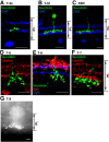
ON cone bipolar cell subtypes were identified by immunohistochemistry after physiological recordings. The IPLs were marked using immunolabeling with calretinin or ChAT antibody. A, Subtype 5s ON bipolar cells (neurobiotin, green) identified with ChAT (blue) immunolabeling. B, Subtype 5f ON bipolar cell. The axon terminal was significantly wider than those of subtype 5s (p < 0.01, n = 7 for subtype 5s, n = 9 for subtype 5f). C, An XBC cell with significantly wider axon terminals was noted next to the inner ChAT band (blue). D, Subtype 6 ON bipolar cell. E, Subtype 6 cells (green) were colabeled with Syt2 (red); ChAT bands (blue). F, Subtype 7 ON bipolar cell. G, Subtype 8 cell labeled with sulforhodamine B, which was identified immediately after physiological recordings. Scale bars, 10 μm.
Figure 2.
Figure 2.
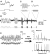
Experimental protocol for step-pulse stimulation and sinusoidal light stimuli. A, The retinal preparation was light adapted at the level of rod saturation (Materials and Methods). A step-pulse light stimulation at 30% contrast was applied every 60 s. Time course of a step-pulse (bottom) and sample L-EPSP trace (top). B, At the same adaptation level, sinusoidal light stimuli of 0.3, 1, 3, 6, 10, and 20 Hz were presented sequentially. C, A sample trace of 6 Hz light stimuli. D, A sample trace of 20 Hz light stimuli. E, A combination of sinusoidal light stimuli (0.15, 0.6, 1, 2.5, 6, 9, 15, and 21 Hz; bottom trace) and the response in an ON bipolar cell (top). FFT analysis revealed the temporal frequencies of sinusoidal stimuli (bottom) and voltage responses (top). The voltage responses evoked in this method were similar to the responses elicited by individual sinusoidal light stimuli.
Figure 3.
Figure 3.
L-EPSPs were increased when stimulus contrast was increased; however, the temporal features did not change. A, When stimulus contrast was increased, L-EPSPs were increased at most of the stimulus frequencies in a transient ON bipolar cell (left) and in a sustained ON bipolar cell (right). B, The L-EPSP amplitude at a peak frequency was plotted as a function of the stimulus contrast levels. C, Higher-contrast sinusoidal stimuli evoked higher L-EPSPs in 7 ON bipolar cells. The amplitude of step light-evoked L-EPSPs were increased when contrast was increased in this range.
Figure 4.
Figure 4.
EPSPs elicited by sinusoidal light stimuli were ON bipolar cell subtype specific. A, Subtype 5s ON bipolar cells were near-low-pass filtering (n = 8, blue curves), whereas subtype 5f cells were bandpass filtering (n = 7, black curves). B, XBCs were bandpass filtering (n = 6). C, Subtype 6 cells were bandpass filtering (n = 7). D, Subtype 7 cells were bandpass filtering with narrow bandwidths. A group of cells exhibited particularly narrow bandwidths compared with another subset of subtype 7 cells (p < 0.05); however, no differences were found between these groups in terms of transient/sustained ratio, high cutoff, or offshoot amplitude. The former group of cells is plotted in red (n = 4), whereas the latter group of cells is shown in black (n = 4). E, Subtype 8 cells were low-pass filtering (n = 5).
Figure 5.
Figure 5.
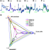
Transient and sustained responses to step-pulse light stimuli were also ON bipolar cell subtype specific. A–F, Step-pulse light stimuli of 30% contrast evoked L-EPSPs in subtype 5s (A), subtype 5f (B), XBC (C), subtype 6 (D), subtype 7 (E), and subtype 8 ON bipolar cells (F). Three to six traces were overlaid from a representative cell in each subtype. G, A summary plot shows three temporal analysis parameters for 6 ON bipolar cell subtypes. For all parameters, temporal tuning is higher from the rear to the front of each axis. Subtypes in a circle were not significantly different from each other. All parameters in subtype 7 cells were the highest, indicating that these cells were highly tuned to a temporal frequency. Conversely, subtype 8 cells were the most sustained cells.
Figure 6.
Figure 6.
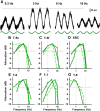
L-EPSCs were bandpass filtering in ON bipolar cells. A, An example of sinusoidal light stimuli-evoked EPSCs. Voltage-clamp mode recordings reflected photoreceptor inputs without voltage-gated channel activation in recorded cells. The L-EPSC amplitude peaked at 3 Hz. BG, L-EPSCs recorded in individual ON bipolar cell are shown in dark green. The average plots of L-EPSPs for each subtype are overlaid (dashed light green).
Figure 7.
Figure 7.
Responses to current injection were low-pass filtering in ON bipolar cells. A, Sinusoidal current injection evoked responses, which were reduced as the frequency increased. B, Sinusoidal current-evoked responses were always low-pass filtering (n = 7). C, Subtype 6 ON bipolar cells. L-EPSPs were bandpass filtering (green), whereas current responses were low-pass filtering (black) in the same cells. D, Subtype 7 ON bipolar cells. Similarly, L-EPSPs (green) were bandpass filtering, whereas responses to current injection (black) were low-pass filtering in the same cells.
Figure 8.
Figure 8.
Summary diagram of the ON cone bipolar cell contribution to temporal processing. ON cone bipolar cells encode distinct temporal visual signaling in a subtype-dependent manner. In most of the IPL sublamina b, both high and low temporal bipolar cells provide synaptic outputs. Subtypes 7 and XBC are especially highly tuned to particular frequencies that provide synaptic outputs near the ChAT band.

References

    1. Armstrong-Gold CE, Rieke F. Bandpass filtering at the rod to second-order cell synapse in salamander (Ambystoma tigrinum) retina. J Neurosci. 2003;23:3796–3806. - PMC - PubMed
    1. Awatramani GB, Slaughter MM. Origin of transient and sustained responses in ganglion cells of the retina. J Neurosci. 2000;20:7087–7095. - PMC - PubMed
    1. Baden T, Esposti F, Nikolaev A, Lagnado L. Spikes in retinal bipolar cells phase-lock to visual stimuli with millisecond precision. Curr Biol. 2011;21:1859–1869. doi: 10.1016/j.cub.2011.09.042. - DOI - PMC - PubMed
    1. Baden T, Berens P, Bethge M, Euler T. Spikes in mammalian bipolar cells support temporal layering of the inner retina. Curr Biol. 2013;23:48–52. doi: 10.1016/j.cub.2012.11.006. - DOI - PubMed
    1. Berntson A, Taylor WR. Response characteristics and receptive field widths of on-bipolar cells in the mouse retina. J Physiol. 2000;524:879–889. doi: 10.1111/j.1469-7793.2000.00879.x. - DOI - PMC - PubMed

Publication types