. 2014:2014:403515.
doi: 10.1155/2014/403515. Epub 2014 May 21.

Porcine reproductive and respiratory syndrome virus induces IL-1β production depending on TLR4/MyD88 pathway and NLRP3 inflammasome in primary porcine alveolar macrophages

Affiliations

Porcine reproductive and respiratory syndrome virus induces IL-1β production depending on TLR4/MyD88 pathway and NLRP3 inflammasome in primary porcine alveolar macrophages

Jing Bi et al. Mediators Inflamm. 2014.

Abstract

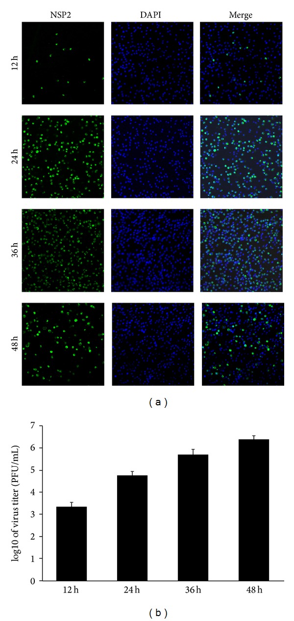
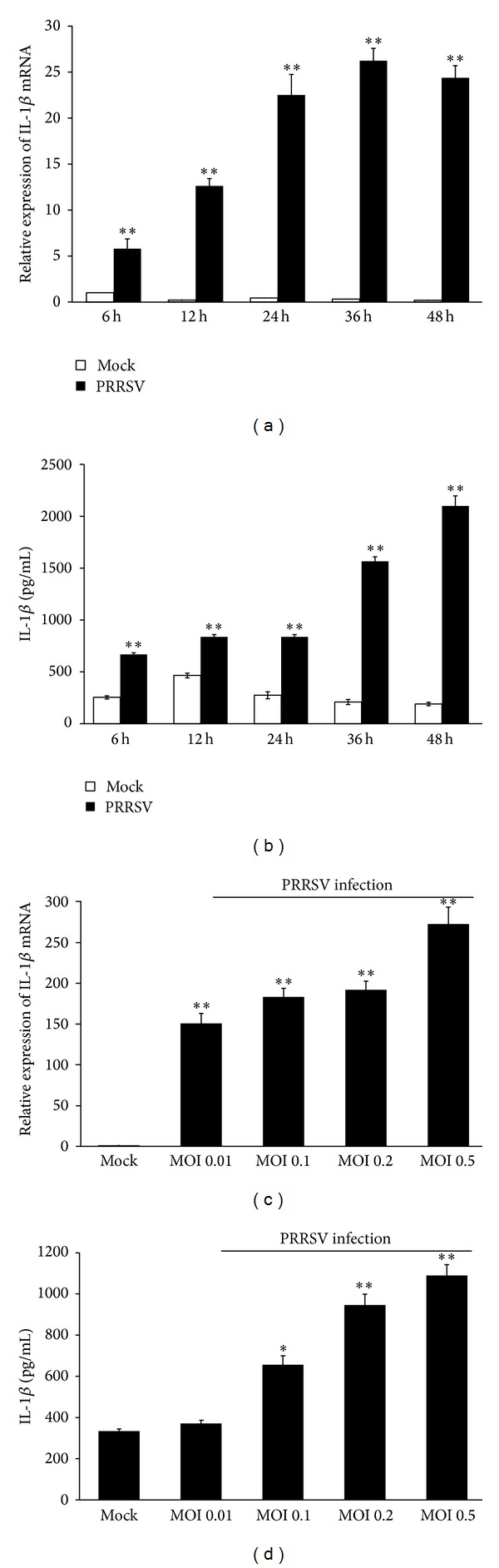
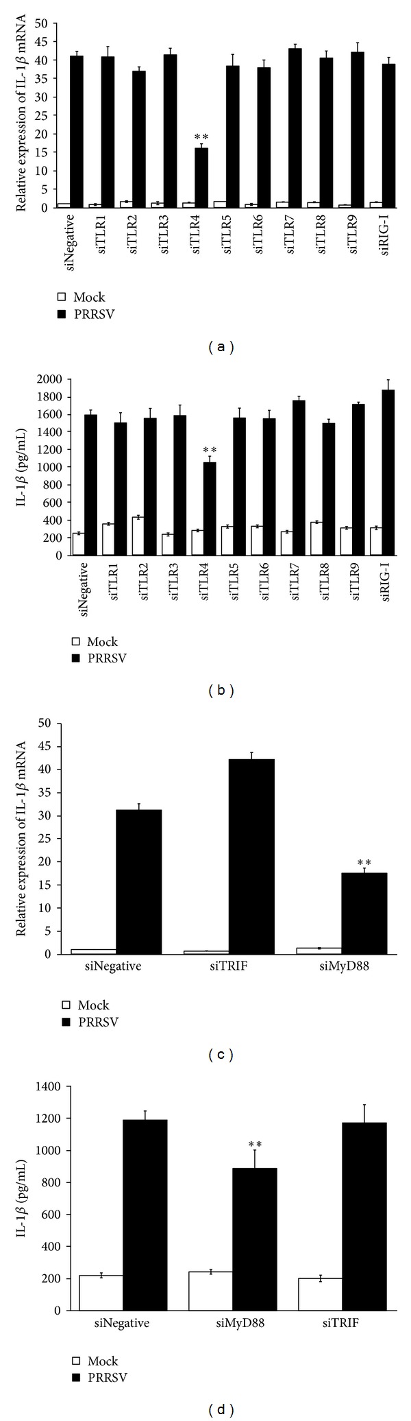
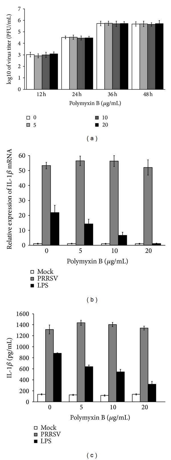
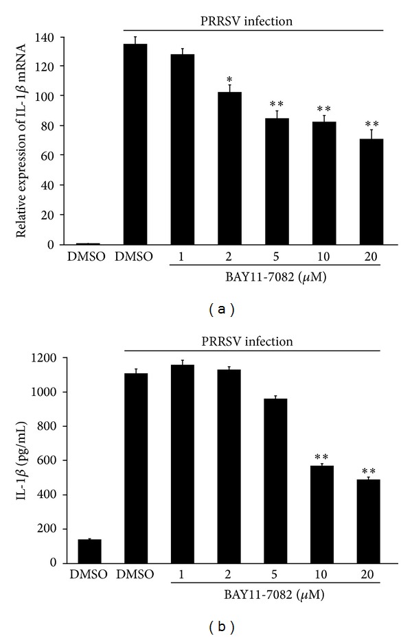
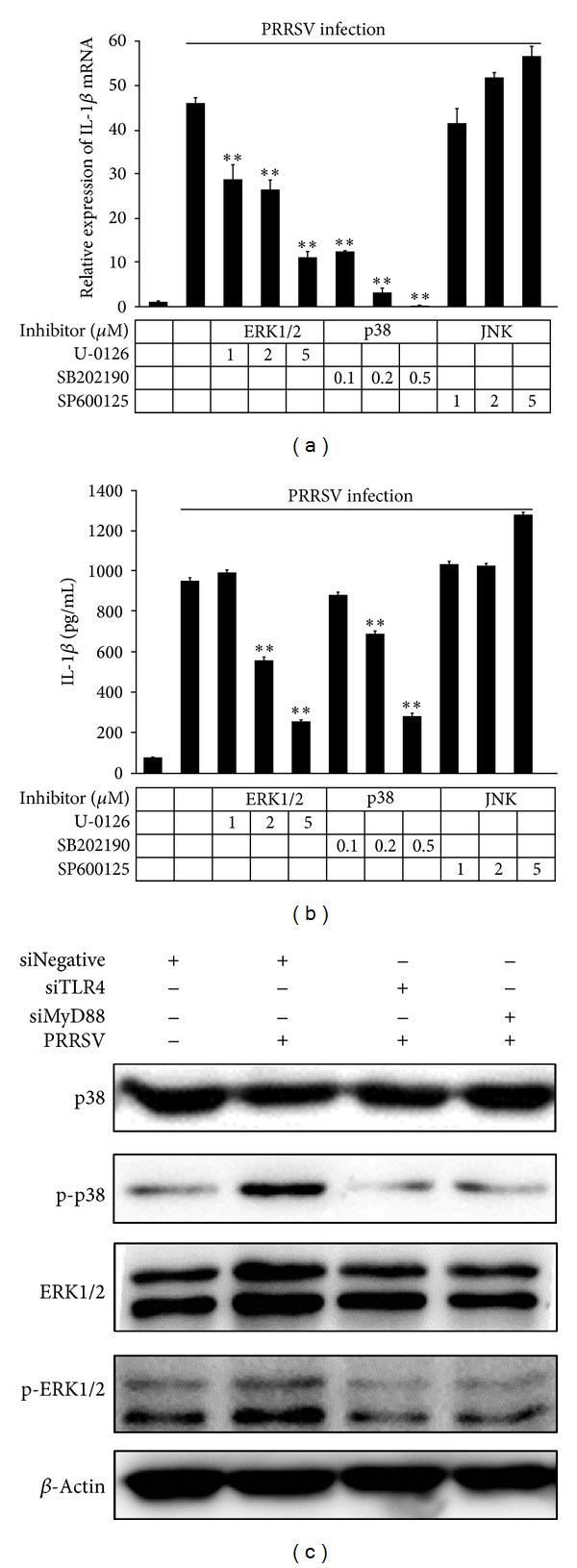
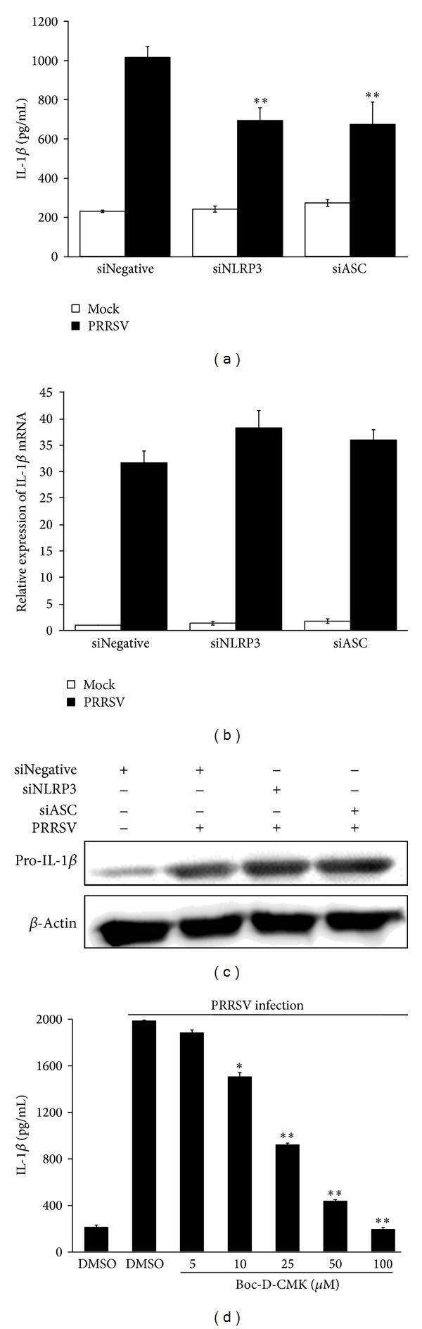
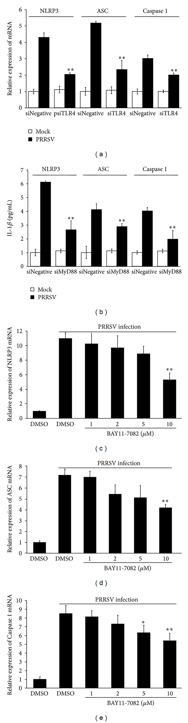
Porcine reproductive and respiratory syndrome virus (PRRSV) is an Arterivirus that has been devastating the swine industry worldwide since the late 1980s. Previous studies have reported that PRRSV infection induced the production of IL-1 β . However, the cellular sensors and signaling pathways involved in this process have not been elucidated yet. Here, we studied the mechanisms responsible for the production of IL-1 β in response to highly pathogenic PRRSV. Upon PRRSV infection of primary porcine alveolar macrophages, both mRNA expression and secretion of IL-1 β were significantly increased in a time- and dose-dependent manner. We also investigated the role of several pattern-recognition receptors and adaptor molecules in this response and showed that the TLR4/MyD88 pathway and its downstream signaling molecules, NF- κ B, ERK1/2, and p38 MAPKs, were involved in IL-1 β production during PRRSV infection. Treatment with specific inhibitors or siRNA knockdown assays demonstrated that components of the NLRP3 inflammasome were crucial for IL-1 β secretion but not for IL-1 β mRNA expression. Furthermore, TLR4/MyD88/NF- κ B signaling pathway was involved in PRRSV-induced expression of NLRP3 inflammasome components. Together, our results deciphered the pathways leading from recognition of PRRSV to the production and release of IL-1 β , providing a deeper knowledge of the mechanisms of PRRSV-induced inflammation responses.


Figures

Figure 1
Figure 1
Infection kinetics of the highly pathogenic PRRSV strain WUH3 in PAMs. (a) PAMs were infected with the adapted PRRSV (3rd passages) at an MOI of 0.1. Cells were fixed and permeabilized in cold methanol at different time point (12, 24, 36, and 48 h) postinfection. Immunofluorescence assays were performed to analyze the replication of PRRSV by detecting the nonstructural protein Nsp2 (green fluorescence). DAPI (4′,6-diamidino-2-phenylindole) was used to stain the nuclei. (b) PAMs were infected with the adapted PRRSV at a MOI of 0.1. Supernatants were collected at different time point (12, 24, 36, and 48 h) postinfection for plaque assay to determine viral titers.
Figure 2
Figure 2
PRRSV infection increases IL-1β mRNA expression and secretion in PAMs. (a, b) PAMs were mock infected or infected with PRRSV at a MOI of 0.1. Cells and supernatants were collected separately at the indicated time points and subjected to real-time RT-PCR (a) and ELISA (b) to analyze the expression of IL-1β. (c, d) PAMs were infected with PRRSV at different doses (0.01, 0.1, 0.2, and 0.5 MOI). Cells and supernatants were collected separately at 36 h postinfection for analysis by IL-1β-specific real-time RT-PCR (c) and ELISA (d). The mock-infected cells were used as negative controls. *P < 0.05 and **P < 0.01 compared with the mock-infected cells.
Figure 3
Figure 3
PRRSV-induced IL-1β production depends on TLR4-MyD88 pathway. (a, b) PAMs were transfected with 80 nM of psiNegative, psiRIG-I, and psiTLR1-9, respectively. At 24 h after transfection, cells were mock infected or infected with PRRSV at a MOI of 0.1. The cells and supernatants were harvested separately 36 h later and subjected to porcine IL-1β-specific real-time RT-PCR (a) and ELISA (b), respectively. (c, d) PAMs were transfected with 80 nM of psiMyD88 and psiTRIF, respectively. At 24 h after transfection, cells were mock infected or infected with PRRSV at a MOI of 0.1. The cells and supernatants were harvested separately 36 h later and analyzed by IL-1β-specific real-time RT-PCR (c) and ELISA (d), respectively. *P < 0.05  and **P < 0.01 compared with cells transfected with psiNegative followed by PRRSV infection.
Figure 4
Figure 4
PMB has no effect on PRRSV proliferation or PRRSV-induced IL-1β production. (a) PAMs were infected with the adapted PRRSV at a MOI of 0.1, followed by treatment with polymyxin B (0, 5, 10, and 20 μg/mL) in the absence of serum, for 36 h. Supernatants were collected at different time point (12, 24, 36, and 48 h) postinfection for plaque assay to determine viral titers. (b, c) PAMs were infected with the adapted PRRSV at a MOI of 0.1 or treated with LPS (1 μg/mL), followed by treatment with polymyxin B (0, 5, 10, and 20 μg/mL) in the absence of serum, for 36 h. The cells and supernatants were then harvested separately and analyzed by real-time RT-PCR and ELISA, respectively.
Figure 5
Figure 5
NF-κB is required for PRRSV-induced IL-1β production. (a, b) PAMs were mock infected or infected with PRSSV at a MOI of 0.1, followed by treatment with NF-κB inhibitor (1, 2, 5, 10, and 20 μM) or DMSO vehicle in the absence of serum, for 36 h. The cells and supernatants were then harvested separately and analyzed by real-time RT-PCR (a) and ELISA (b), respectively. *P < 0.05 and **P < 0.01 compared with DMSO-treated cells plus PRRSV infection.
Figure 6
Figure 6
MAPK p38 and ERK1/2 are critical for PRRSV-induced IL-1β production in PAMs. Experiments were performed similar to those described in Figure 3 except that ERK1/2 inhibitor (1, 2, and 5 μM), p38 MAPK inhibitor (0.1, 0.2, and 0.5 μM), or JNK inhibitor (1, 2, and 5 μM) was used. Cells and supernatants were then harvested and analyzed by IL-1β-specific real-time RT-PCR (a) and ELISA (b), respectively. **P < 0.01 compared with DMSO plus PRRSV infection.
Figure 7
Figure 7
PRRSV-induced IL-1β secretion requires NLRP3 inflammasome. (a–c) PAMs were transfected with 80 nM of psiNegative, psiNLRP3, or psiASC, respectively. At 24 h posttransfection, cells were mock infected or infected with PRRSV at a MOI of 0.1. The supernatants and cells were harvested separately 36 h later and analyzed by IL-1β ELISA (a) and real-time RT-PCR (b), respectively. For Western blotting to detect pro-IL-1β, cells were harvested at 24 h postinfection by using polyclonal antibody against pro-IL-1β (c). (d) PAMs were mock infected or infected with PRRSV at a MOI of 0.1, followed by treatment with caspase 1 inhibitor (5, 10, 25, 50, and 100 μM) or DMSO vehicle in the absence of serum, for 36 h. The supernatants were then harvested and analyzed by IL-1β-specific ELISA. *P < 0.05 and **P < 0.01 compared with DMSO-treated cells plus PRRSV infection.
Figure 8
Figure 8
TLR4/MyD88/NF-κB signaling pathway is involved in PRRSV-induced expression of NLRP3 inflammasome components. (a, b) PAMs were transfected with 80 nM of psiNegative, psiTLR4, or psiMyD88, respectively. At 24 h posttransfection, cells were mock infected or infected with PRRSV at a MOI of 0.1. The cells and supernatants were harvested separately 36 h later and analyzed by NLRP3/ASC/caspase 1 specific real-time RT-PCR. (c–e) PAMs were mock infected or infected with PRSSV at a MOI of 0.1, followed by treatment with NF-κB inhibitor (1, 2, 5, and 10 μM) or DMSO vehicle in the absence of serum, for 36 h. The cells were then harvested to analyse the mRNA expression of NLRP3 (c), ASC (d), and caspase 1 (e), respectively, by real-time RT-PCR. *P < 0.05 and **P < 0.01 compared with DMSO-treated cells plus PRRSV infection.

Similar articles

Cited by

References

    1. Bose S, Banerjee AK. Innate immune response against nonsegmented negative strand RNA viruses. Journal of Interferon and Cytokine Research. 2003;23(8):401–412. - PubMed
    1. Dinarello CA. Biologic basis for Interleukin-1 in disease. Blood. 1996;87(6):2095–2147. - PubMed
    1. Eder C. Mechanisms of interleukin-1β release. Immunobiology. 2009;214(7):543–553. - PubMed
    1. Lopez-Castejon G, Brough D. Understanding the mechanism of IL-1β secretion. Cytokine and Growth Factor Reviews. 2011;22(4):189–195. - PMC - PubMed
    1. Takeuchi O, Akira S. Pattern recognition receptors and inflammation. Cell. 2010;140(6):805–820. - PubMed

Publication types

MeSH terms