. 2014:2014:935251.
doi: 10.1155/2014/935251. Epub 2014 May 21.

Hydrogen sulfide ameliorates ischemia/reperfusion-induced hepatitis by inhibiting apoptosis and autophagy pathways

Affiliations

Hydrogen sulfide ameliorates ischemia/reperfusion-induced hepatitis by inhibiting apoptosis and autophagy pathways

Ping Cheng et al. Mediators Inflamm. 2014.

Abstract

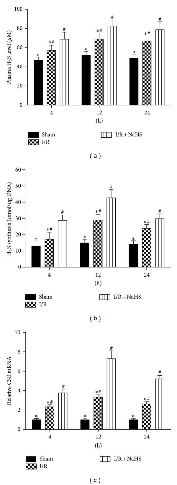
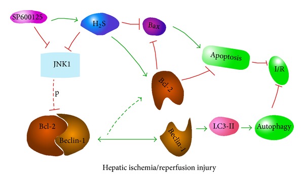
Background: Hepatic ischemia/reperfusion (I/R) injury is an important clinical problem, and its consequences can seriously threaten human health. Apoptosis and autophagy have been shown to contribute to cell death in hepatic I/R injury. Hydrogen sulfide (H2S) is the third most common endogenously produced gaseous signaling molecule and is known to exert a protective effect against hepatic I/R injury. In this study, the purpose is to explore both the effect and mechanism of H2S on hepatic I/R injury.

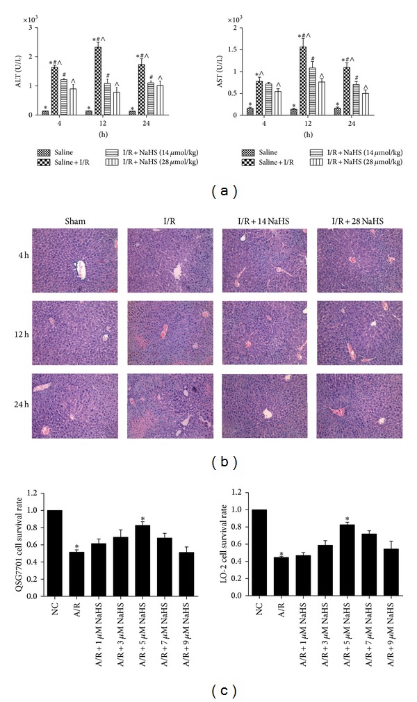
Methods: Balb/c mice were randomized into Sham, I/R, or two doses (14 μmol/kg and 28 μmol/kg) of sodium hydrosulfide (NaHS, an H2S donor) preconditioning groups.

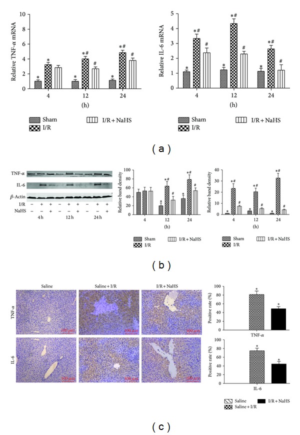
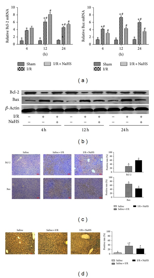
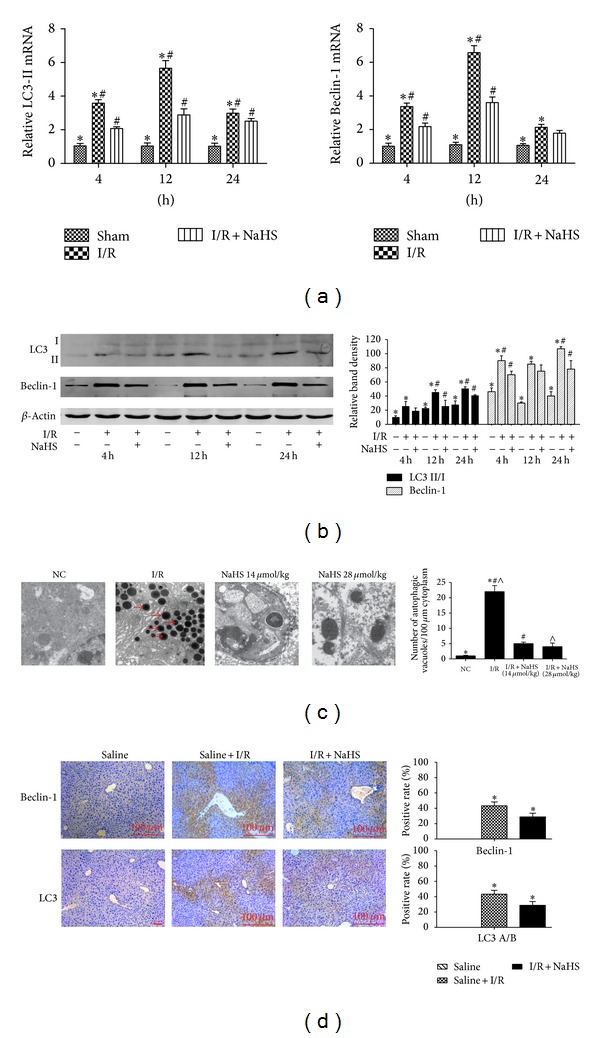
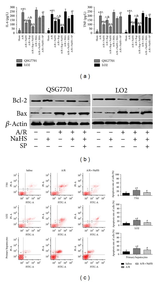
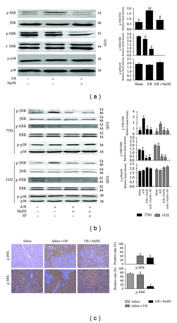
Results: NaHS significantly reduced the levels of TNF- α and IL-6 at 12 h and 24 h after injection compared with ischemia/reperfusion challenge alone. The expression of Bcl-2, Bax, Beclin-1, and LC3, which play important roles in the regulation of the apoptosis and autophagy pathways, was also clearly affected by NaHS. Furthermore, NaHS affected the p-JNK1, p-ERK1, and p-p38.

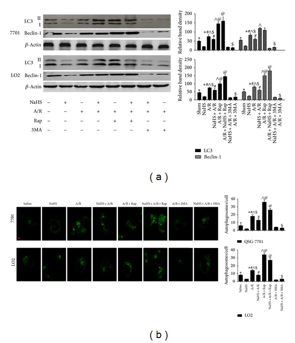
Conclusion: Our results indicate that H2S attenuates hepatic I/R injury, at least in part, by regulating apoptosis through inhibiting JNK1 signaling. The autophagy agonist rapamycin potentiated this hepatoprotective effect by reversing the inhibition of autophagy by H2S.


Figures

Figure 1
Figure 1
Plasma H2S level, H2S-synthesizing activity, and CSE mRNA expression in livers. (a) Plasma hydrogen sulfide (H2S) concentration was expressed as the mean ± SD of 6 animals per group. *P < 0.05 for saline versus saline + I/R, # P < 0.05 for saline + I/R versus I/R + NaHS (14 μmol/kg). (b) Liver H2S-synthesizing activity was expressed as the mean ± SD of 6 animals per group. *P < 0.05 for saline versus saline + I/R, # P < 0.05 for saline + I/R versus I/R + NaHS (14 μmol/kg). (c) The mRNA expression of CSE was detected by real time PCR. *P < 0.05 for saline versus saline + I/R, # P < 0.05 for saline + I/R versus I/R + NaHS (14 μm/kg).
Figure 2
Figure 2
Effect of NaHS on hepatic ischemia-reperfusion injury. (a) The serum ALT and AST levels were expressed as the mean ± SD of 6 animals per group. *P < 0.05 for saline versus saline + I/R, # P < 0.05 for saline + I/R versus I/R + NaHS (14 μmol/kg), and P < 0.05 for saline + I/R versus I/R + NaHS (28 μmol/kg). (b) Representative hematoxylin and eosin (H&E) stained sections of liver. Original magnifications: ×200. (c) Hepatocytes (LO2, QSG7701) that were subjected to NaHS (1 μM, 3 μM, 5 μM, 7 μM, and 9 μM), then the cells were treated with 24 h hypoxia (3% O2, 5% CO2, and 92% N2) and 2 h reoxygenation (5% CO2, 95% air). Cell viability was measured using an MTT assay and a microplate reader at a wavelength of 490 nm. The experiments were repeated three times. The results are expressed as the mean ± SD (n = 5). *P < 0.05 for (A/R + 5 μM NaHS) versus saline + A/R.
Figure 3
Figure 3
NaHS pretreatment inhibits the release of cytokines during hepatic ischemia-reperfusion (I/R) injury. (a) The mRNA expression of IL-6 and TNF-α in liver tissues was detected by real time PCR. *P < 0.05 for saline versus saline + I/R, # P < 0.05 for saline + I/R versus I/R + NaHS (14 μmol/kg). (b) Western blots and quantitative evaluation of the expression of IL-6 and TNF-α in liver tissues with β-actin as protein loading control. *P < 0.05 for saline versus saline + I/R, # P < 0.05 for saline + I/R versus I/R + NaHS (14 μmol/kg). (c) Immunohistochemistry staining (200x) showed the expression of TNF-α and IL-6 in liver tissue at 12 h. *P < 0.05 for (saline + I/R) versus (I/R + NaHS).
Figure 4
Figure 4
Effect of NaHS pretreatment on regulation of apoptosis in vivo. (a) The mRNA expression of Bcl-2 and Bax in liver tissues was detected by real time PCR. *P < 0.05 for saline versus saline + I/R, # P < 0.05 for saline + I/R versus I/R + NaHS (14 μmol/kg). (b) Western blots and quantitative evaluation of the expression of Bcl-2 and Bax in liver tissues. *P < 0.05 for saline versus saline + I/R,# P < 0.05 for saline + I/R versus I/R + NaHS (14 μmol/kg). (c) Immunohistochemistry staining (200x) showed the expression of Bcl-2 and Bax in liver tissue at 12 h. *P < 0.05 for saline + I/R versus I/R + NaHS. (d) TUNNEL staining showed the apoptotic cells in three groups at 12 h. Original magnifications: ×200. *P < 0.05 for saline versus saline + I/R, # P < 0.05 for saline + I/R versus I/R + NaHS.
Figure 5
Figure 5
Effect of NaHS pretreatment on regulation of autophagy in vivo. (a) The mRNA expression of Beclin-1 and LC3 in liver tissues was detected by real time PCR. *P < 0.05 for saline versus saline + I/R, # P < 0.05 for saline + I/R versus I/R + NaHS (14 μmol/kg). (b) Western blots and quantitative evaluation of the expression of Beclin-1 and LC3 in liver tissues with β-actin as protein loading control. *P < 0.05 for saline versus saline + I/R, # P < 0.05 for saline + I/R versus I/R + NaHS (14 um/kg). (c) Electron microscopy showed the ultrastructure and autophagosomes (“→” indicated the autophagosomes). Original magnifications: ×2500. *P < 0.05 for saline versus saline + I/R, #, ∧ P < 0.05 for saline + I/R versus I/R + NaHS. (d) Immunohistochemistry staining (200x) showed the expression of Beclin-1 and LC3 protein in liver tissue at 12 h. *P < 0.05 for saline + I/R versus I/R + NaHS.
Figure 6
Figure 6
Effect of NaHS pretreatment on regulation of apoptosis in vitro. (a) IL-6 and TNF levels were assessed by ELISA. Data are expressed as mean ± SD of 5 wells nutrient solution per group. *Significant difference from control, P < 0.05; #significant difference from A/R group, P < 0.05; P < 0.05 for A/R versus A/R + Rap; $ P < 0.05 for A/R + NaHS versus A/R + NaHS + Rap; @ P < 0.05 for A/R + NaHS versus A/R + NaHS + SP. (b) Western blots of the expression of Bcl-2 and Bax in vitro that were subjected to saline, A/R, A/R + NaHS, and A/R + NaHS + SP with β-actin as protein loading control. (c) Flow cytometric analyses of annexin-V/PI staining of hepatocytes (LO2, QSG7701, and primary hepatocytes). *P < 0.05 for saline versus saline + A/R, # P < 0.05 for saline + A/R versus A/R + NaHS.
Figure 7
Figure 7
Effect of NaHS pretreatment on regulation of autophagy in vitro. (a) Western blots and quantitative evaluation of the expression of Beclin-1 and LC3 in vitro with β-actin as protein loading control. *Significant difference from control, P < 0.05; #significant difference from A/R group, P < 0.05; P < 0.05 for A/R versus A/R + Rap; $ P < 0.05 for A/R + NaHS versus A/R + NaHS + Rap; @ P < 0.05 for A/R + NaHS versus A/R + NaHS + SP. (b) The average number of autophagosomes/cell ± SD counted from confocal microscopy images of hepatocytes (LO2, QSG7701) expressing GFP-LC3. (Original magnifications: ×400.)
Figure 8
Figure 8
The effect of JNK1 in the protective effect of H2S on hepatocyte A/R and I/R injuries. (a) Western blots and quantitative evaluation of the expression of p-JNK, p-ERK, and p-p38 in vivo. *P < 0.05 for saline versus saline + I/R, # P < 0.05 for saline + I/R versus I/R + NaHS. (b) Western blots and quantitative evaluation of the expression of p-JNK, p-ERK, and p-p38 in vitro. *P < 0.05. (c) Immunohistochemistry staining (200x) showed the expression of p-JNK and p-ERK in liver tissue at 12 h. *P < 0.05 for saline + I/R versus I/R + NaHS.
Figure 9
Figure 9
In the condition of hepatic ischemia-reperfusion injury, H2S reduces autophagy (which is an important protective mechanism against I/R- and A/R-induced hepatitis) through the suppression of the JNK pathway. However, it also plays an antiapoptotic role in ameliorating I/R- and A/R-induced hepatitis and these protective effects are enhanced by the inhibition of JNK. Our findings show that, although autophagy was inhibited by H2S, H2S still showed a protective effect against I/R. Furthermore, the JNK1-mediated phosphorylation of Bcl-2 substantially reduced the affinity of Bcl-2 for Beclin-1, leading to its rapid dissociation from Beclin-1 and the subsequent induction of prosurvival autophagy, reducing hepatic I/R injury. In our study, the reduced phosphorylation of JNK1 (compared with that in the I/R and A/R groups) by preconditioning with NaHS may have weakened the process described above.

Similar articles

Cited by

References

    1. Dogan S, Aslan M. Hepatic ischemia-reperfusion injury and therapeutic strategies to alleviate cellular damage. Hepatology Research. 2011;41(2):103–117. - PubMed
    1. Chen M, Du Y, Qui M, et al. Ophiopogonin B-induced autophagy in non-small cell lung cancer cells via inhibition of the PI3K/Akt signaling pathway. Oncology Reports. 2013;29(2):430–436. - PMC - PubMed
    1. Tang G, Yue Z, Talloczy Z, et al. Autophagy induced by Alexander disease-mutant GFAP accumulation is regulated by p38/MAPK and mTOR signaling pathways. Human Molecular Genetics. 2008;17(11):1540–1555. - PMC - PubMed
    1. Shen M, Lu J, Dai W, et al. Ethyl pyruvate ameliorates hepatic ischemia-reperfusion injury by inhibiting intrinsic pathway of apoptosis and autophagy. Mediators of Inflammation. 2013;2013:12 pages.461536 - PMC - PubMed
    1. Shi J, Li XH, Sun BC, et al. Protective functions of recombinant protein targeted at RANKL against hepatic ischemia/reperfusion injury transfected by retrovirus in mice. Zhonghua Yi Xue Za Zhi. 2011;91(38):2719–2724. - PubMed

Publication types

MeSH terms