Review
. 2014 Jul;29(3):269-78.
doi: 10.1007/s12291-013-0408-y. Epub 2013 Nov 26.

Alkaline phosphatase: an overview

Affiliations
Review

Alkaline phosphatase: an overview

Ujjawal Sharma et al. Indian J Clin Biochem. 2014 Jul.

Abstract

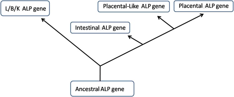
Alkaline phosphatase (ALP; E.C.3.I.3.1.) is an ubiquitous membrane-bound glycoprotein that catalyzes the hydrolysis of phosphate monoesters at basic pH values. Alkaline phosphatase is divided into four isozymes depending upon the site of tissue expression that are Intestinal ALP, Placental ALP, Germ cell ALP and tissue nonspecific alkaline phosphatase or liver/bone/kidney (L/B/K) ALP. The intestinal and placental ALP loci are located near the end of long arm of chromosome 2 and L/B/K ALP is located near the end of the short arm of chromosome 1. Although ALPs are present in many mammalian tissues and have been studied for the last several years still little is known about them. The bone isoenzyme may be involved in mammalian bone calcification and the intestinal isoenzyme is thought to play a role in the transport of phosphate into epithelial cells of the intestine. In this review, we tried to provide an overview about the various forms, structure and functions of alkaline phosphatase with special focus on liver/bone/kidney alkaline phosphatase.

Keywords: Alkaline phosphatase; Enzymes; Intestinal alkaline phosphatase; Isoenzymes; L/B/K alkaline phosphatase; Liver alkaline phosphatase; Placental alkaline phosphatase.


Figures

Fig. 1
Fig. 1
Illustration showing the postulated evolutionary relationships of the human liver/bone/kidney, intestinal, placental and placental-like genes [18]
Fig. 2
Fig. 2
Relationship between exon organization and polypeptide structure of the L/B/K ALP gene. L/B/K ALP gene exons 1–12 are shown as large rectangles. Untranslated regions are indicated by green colour. The signal peptide at the amino terminus and the hydrophobic stretch of amino acids at the carboxyl terminus in exons 2 and 12, respectively, are shown in yellow. Regions which comprise the active pocket that are conserved in intestinal ALP, placental ALP, and E. coli ALP are shown as follows: small rectangles above the exons indicate conserved units of amino acid sequence which exist as discrete units of secondary structure in E. coli ALP (black for & sheets, white for a-helices); the open circles indicate metal ligands, and the closed circles indicate residues that directly interact with incoming substrate
Fig. 3
Fig. 3
DNA sequence and deduced amino acid sequence of the L/B/K ALP cDNA. Numbers preceded by + or–refer to amino acid positions. All other numbers refer to nucleotide positions. Asterisks occur at 10-base intervals. Amino acids -17 to -1 comprise a putative signal peptide. A vertical line precedes amino acid +1, the amino-terminal residue found in the mature protein. Amino acid residues that have been determined by protein sequence analysis of purified liver ALP are underlined. Five potential N-linked glycosylation signals, Asn-Xaa-Thr/Ser, are boxed. A 12-bp direct repeat in the 3′ untranslated region of the cDNA is labeled by arrows. A single poly(A) addition signal AATAAA is underlined twice [10]
Fig. 4
Fig. 4
Comparison of the amino acid sequences of E. coli (E), human placental (P), and human L/B/K (L) ALP precursor proteins. Gaps that have been introduced into the sequences to maximize pairing of homologous amino acids are indicated by -Amino acid +1 corresponds to the first residue in each of the mature proteins. Amino acids that are identical in all three proteins or in the two human proteins are boxed. Amino acids are shown in the single-letter code [10]
Fig. 5
Fig. 5
A ribbon diagram of L/B/K ALP protein structure. (http://biochem.dental.upenn.edu/)

References

    1. McComb RB, Bowers GN, Posen S. Alkaline phosphatase. New York: Plenum Publishing Corp; 1979.
    1. Tsai LC, Hung MW, Chen YH, Su WC, Chang GG, Chang TC. Expression and regulation of alkaline phosphatases in human breast cancer MCF-7 cells. Eur J Biochem. 2000;267:1330–1339. doi: 10.1046/j.1432-1327.2000.01100.x. - DOI - PubMed
    1. Chang TC, Wang JK, Hung MW, Chiao CH, Tsai LC, Chang GG. Regulation of the expression of alkaline phosphatase in a human breast cancer cell line. Biochem J. 1994;303:199–205. - PMC - PubMed
    1. Whyte MP, Landt M, Ryan LM, Mulivor RA, Henthorn PS, Fedde KN, Mahuren JD, Coburn SP. Alkaline phosphatase: placental and tissue-nonspecific isoenzymes hydrolyze phosphoethanolamine, inorganic pyrophosphate, and pyridoxal 5′-phosphate substrate accumulation in carriers of hypophosphatasia corrects during pregnancy. J Clin Invest. 1995;95:1440–1445. doi: 10.1172/JCI117814. - DOI - PMC - PubMed
    1. Calhau C, Martel F, Hipolito-Reis C, Azevedo I. Effect of P-glycoprotein modulators on alkaline phosphataseactivity in cultured rat hepatocytes. Cell Physiol Biochem. 2000;10:195–202. doi: 10.1159/000016350. - DOI - PubMed

LinkOut - more resources