

Microsurgical training model for residents to approach to the orbit and the optic nerve in fresh cadaveric sheep cranium
- PMID: 24966554
- PMCID: PMC4064181
- DOI: 10.4103/0976-3147.131660
Microsurgical training model for residents to approach to the orbit and the optic nerve in fresh cadaveric sheep cranium
Abstract
Background: Neurosurgery and ophthalmology residents need many years to improve microsurgical skills. Laboratory training models are very important for developing surgical skills before clinical application of microsurgery. A simple simulation model is needed for residents to learn how to handle microsurgical instruments and to perform safe dissection of intracranial or intraorbital nerves, vessels, and other structures.
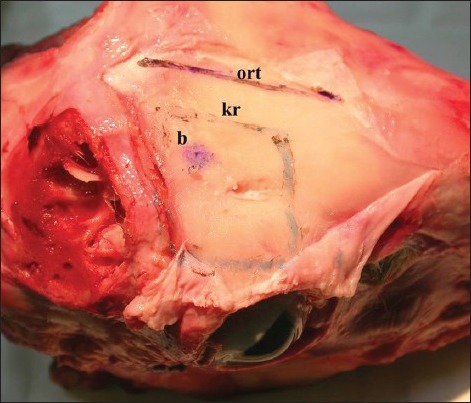
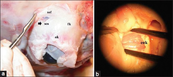
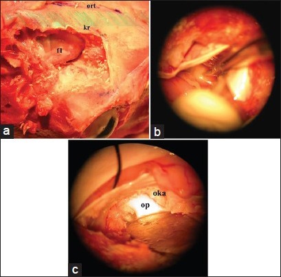
Materials and methods: The simulation material consists of a one-year-old fresh cadaveric sheep cranium. Two parts (Part 1 and Part 2) were designed to approach structures of the orbit. Part 1 consisted of a 2-step approach to dissect intraorbital structures, and Part 2 consisted of a 3-step approach to dissect the optic nerve intracranially.
Results: The model simulates standard microsurgical techniques using a variety of approaches to structures in and around the orbit and the optic nerve.
Conclusions: This laboratory training model enables trainees to gain experience with an operating microscope, microsurgical instruments and orbital structures.
Keywords: Microneurosurgery; microsurgery; microsurgical training; optic nerve; orbita surgery.
Conflict of interest statement
Figures
References
-
- Aboud E, Al-Mefty O, Yaşargil MG. New laboratory model for neurosurgical training that simulates live surgery. J Neurosurg. 2002;97:1367–72. - PubMed
-
- Hicdonmez T, Hamamcioglu MK, Parsak T, Cukur Z, Cobanoglu S. A laboratory training model for interhemispheric-transcallosal approach to the lateral ventricle. Neurosurg Rev. 2006;29:159–62. - PubMed
-
- Hicdonmez T, Hamamcioglu MK, Tiryaki M, Cukur Z, Cobanoglu S. Microneurosurgical training model in fresh cadaveric cow brain: A laboratory study simulating the approach to the circle of Willis. Surg Neurol. 2006;66:100–4. - PubMed
-
- Hicdönmez T, Birgili B, Tiryaki M, Parsak T, Çobanoğlu S. Posterior fossa approach: Microneurosurgical training model in cadaveric sheep. Turk Neurosurg. 2006;16:111–4.
-
- Bao JY. Rat tail: A useful model for microvascular training. Microsurgery. 1995;16:122–5. - PubMed
LinkOut - more resources
Full Text Sources
Other Literature Sources