

doi: 10.4103/0976-3147.131680.
Epidural hematoma with detachment of the dural sinuses
Affiliations
- PMID: 24966568
- PMCID: PMC4064195
- DOI: 10.4103/0976-3147.131680
Item in Clipboard
Epidural hematoma with detachment of the dural sinuses
J Neurosci Rural Pract.
2014 Apr.
Abstract
Epidural hematoma (EH) is a neurosurgical emergency that requires early surgical treatment. It is rarely extended bilaterally causing a detachment of the dural sinus or sinuses. The authors present two rare cases of EH with dural sinus detachment and describe how they suspend them. In these cases it is crucial to firmly suspend the dura mater and the dural sinus to the inner skull surface to prevent postoperative rebleeding.
Keywords: Extradural hematoma; head injuries; operative technique.
Conflict of interest statement
Figures

A computed tomography scan showing a right frontal extradural hematoma with contralateral extension and detachment of the sagittal longitudinal sinus

Illustration (a) of the patient position according to surgeon's view; a bicoronal skin incision flap is traced. Surgical exposure (b) and illustration of the craniotomies after the blood clot evacuation and hemostasis; only the suspensions for the dural sinus are drawn. The dural suspensions (c) are being tied above the strip of bone

A computed tomography scan showing a left cerebellar extradural hematoma. The hematoma extended to the supratentorial space and detached the sinus confluence, the left transverse sinus, and posterior third of the sagittal longitudinal sinus
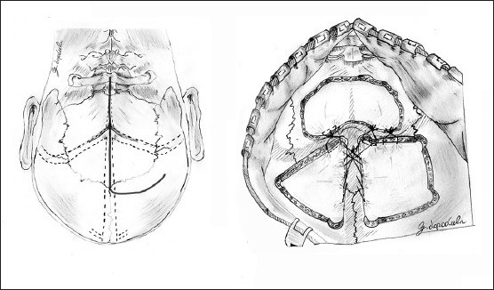
Illustration of the patient position according to surgeon's view, a “hockey stick” skin incision flap is traced (left). Surgical exposure and illustration of the suboccipital craniectomy and of the supratentorial craniotomies after the blood clot evacuation and hemostasis (right). In the sketch, only the suspensions for the dural sinus are drawn
References
-
- Jamieson KG, Yelland JD. Extradural hematoma. Report of 167 cases. J Neurosurg. 1968;29:13–23. - PubMed
-
- McKissock W. Extradural hematoma: Observation in 125 cases. Lancet. 1960;276(7143):167–72. http://dx.doi.org/10.1016/S0140-6736(60) 91322-2 .
-
- Dharker SR, Bhargava N. Bilateral epidural haematoma. Acta Neurochir (Wien) 1991;110:29–32. - PubMed
-
- Frank E, Berger TS, Tew JM., Jr Bilateral epidural hematomas. Surg Neurol. 1982;17:218–22. - PubMed
-
- Roy GC. Fracture of skull, extensive extravasation of blood on dura mater, producing compression of brain, trepanning, partial relief of symptoms, death. Lancet. 1884;2:319.
LinkOut - more resources
Full Text Sources
Other Literature Sources