

Electromyographic, cerebral, and muscle hemodynamic responses during intermittent, isometric contractions of the biceps brachii at three submaximal intensities
- PMID: 24966837
- PMCID: PMC4052733
- DOI: 10.3389/fphys.2014.00190
Electromyographic, cerebral, and muscle hemodynamic responses during intermittent, isometric contractions of the biceps brachii at three submaximal intensities
Abstract
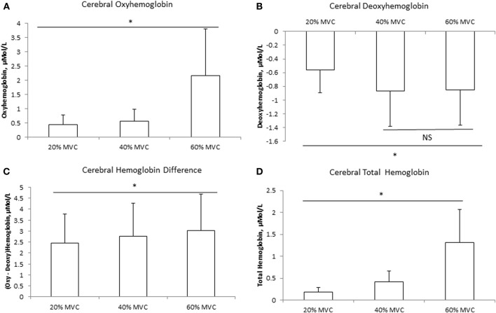
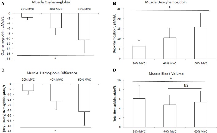
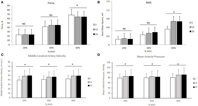
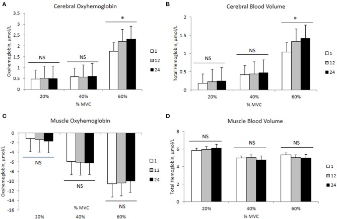
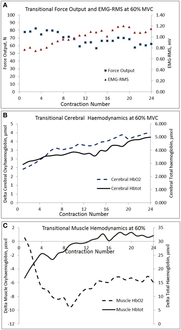
This study examined the electromyographic, cerebral and muscle hemodynamic responses during intermittent isometric contractions of biceps brachii at 20, 40, and 60% of maximal voluntary contraction (MVC). Eleven volunteers completed 2 min of intermittent isometric contractions (12/min) at an elbow angle of 90° interspersed with 3 min rest between intensities in systematic order. Surface electromyography (EMG) was recorded from the right biceps brachii and near infrared spectroscopy (NIRS) was used to simultaneously measure left prefrontal and right biceps brachii oxyhemoglobin (HbO2), deoxyhemoglobin (HHb), and total hemoglobin (Hbtot). Transcranial Doppler ultrasound was used to measure middle cerebral artery velocity (MCAv) bilaterally. Finger photoplethysmography was used to record beat-to-beat blood pressure and heart rate. EMG increased with force output from 20 to 60% MVC (P < 0.05). Cerebral HbO2 and Hbtot increased while HHb decreased during contractions with differences observed between 60% vs. 40% and 20% MVC (P < 0.05). Muscle HbO2 decreased while HHb increased during contractions with differences being observed among intensities (P < 0.05). Muscle Hbtot increased from rest at 20% MVC (P < 0.05), while no further change was observed at 40 and 60% MVC (P > 0.05). MCAv increased from rest to exercise but was not different among intensities (P > 0.05). Force output correlated with the root mean square EMG and changes in muscle HbO2 (P < 0.05), but not changes in cerebral HbO2 (P > 0.05) at all three intensities. Force output declined by 8% from the 1st to the 24th contraction only at 60% MVC and was accompanied by systematic increases in RMS, cerebral HbO2 and Hbtot with a leveling off in muscle HbO2 and Hbtot. These changes were independent of alterations in mean arterial pressure. Since cerebral blood flow and oxygenation were elevated at 60% MVC, we attribute the development of fatigue to reduced muscle oxygen availability rather than impaired central neuronal activation.
Keywords: biceps force; cerebral blood flow and oxygenation; electromyography; muscle blood flow and oxygenation.
Figures
References
-
- Bhambhani Y. (2012). Application of near infrared spectroscopy to evaluate cerebral and muscle hemodynamics during exercise and sport. JNIRS 20, 117–139 10.1255/jnirs.978 - DOI
LinkOut - more resources
Full Text Sources
Other Literature Sources