. 2014 Apr 15;7(5):1889-99.
eCollection 2014.

TSLP induced by estrogen stimulates secretion of MCP-1 and IL-8 and growth of human endometrial stromal cells through JNK and NF-κB signal pathways

Affiliations

TSLP induced by estrogen stimulates secretion of MCP-1 and IL-8 and growth of human endometrial stromal cells through JNK and NF-κB signal pathways

Kai-Kai Chang et al. Int J Clin Exp Pathol. .

Abstract

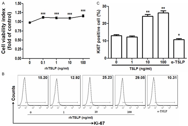
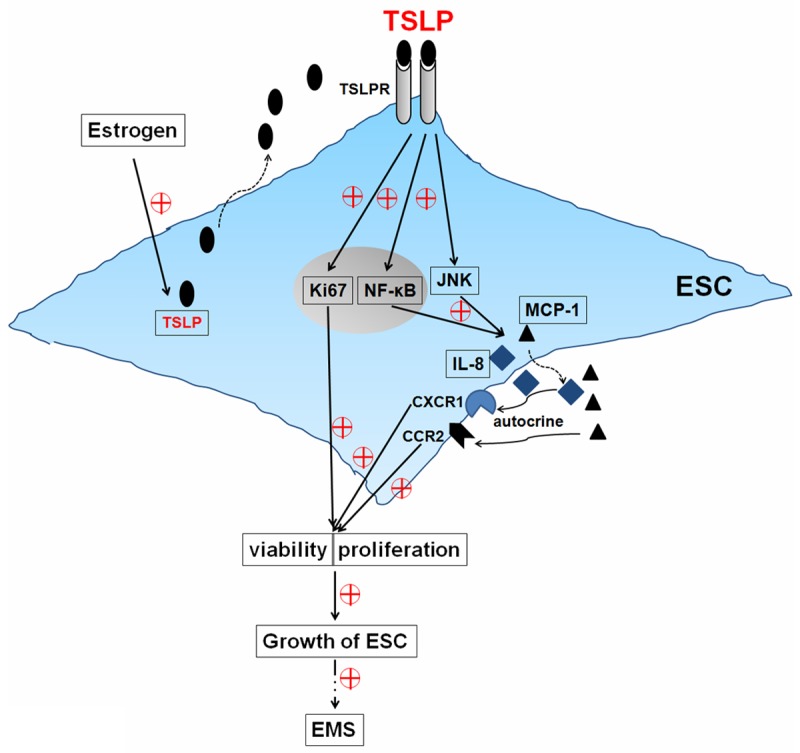
It has reported that human endometrial stromal cells (ESCs) express thymic stromal lymphopoietin (TSLP), and TSLP concentrations in the serum and peritoneal fluid were higher in women with endometriosis. Endometriosis is an estrogen-dependent disease. The present study aimed to elucidate whether and how estrogen regulates the growth of ESCs through TSLP. The ESCs behaviors in vitro were verified by SRB assay and Ki67 level detection, respectively. In addition, the effects of estrogen on TSLP and TSLP on the correspondent functional molecules were investigated by ELISA and flow cytometry. Here we found that estrogen stimulated the secretion of TSLP in a dosage-dependent manner. Recombinant human TSLP stimulates the secretion of MCP-1 and IL-8, and markedly promotes the viability and proliferation relative gene Ki-67 expression of ESCs. These effects could be abolished by the inhibitor for JNK or NF-κB signal, respectively. Moreover, not only anti-TSLP neutralizing antibody, but also blocking JNK or NF-κB signal by inhibitor abrogated the stimulatory role in the production of MCP-1 and IL-8, and the growth of ESCs induced by estrogen. Our current study has demonstrated that TSLP is involved in the regulation of estrogen on the secretion of MCP-1 and IL-8, and the growth of ESCs through JNK and NF-κB signal pathways, which suggests that the abnormal high expression of TSLP induced by estrogen may play an important role in ESCs growth and finally contribute to the origin and development of endometriosis.

Keywords: ESCs; IL-8; MCP-1; TSLP; endometriosis; estrogen; proliferation.


Figures

Figure 1
Figure 1
Estrogen stimulates the secretion of TSLP in ESCs. ESCs (1×10⁵ cells/well) of eutopic endometrium from women with endometriosis (n=6) were seeded in 24-well plates, and treated with 17-β estradiol (10-11 M-10-7 M) for 48 h, with the vehicle as control, and then ELISA was performed to analyze the secretion level of TSLP in the supernatant. E2: 17-β estradiol. Error bars depict the standard deviation of the mean. P<0.05, **P<0.01 and ***P<0.001 compared to control. ##P<0.01 compared to 10-11 M group.
Figure 2
Figure 2
TSLP enhances the viability and proliferation of ESCs. We incubated ESCs (n=6) (1×10⁴ cells/well in 96-well plates for SRB assay; 1×10⁵ cells/well in 96-well plates for flow cytometry) with recombinant human TSLP (rhTSLP) from 0.1 to 100 ng/ml or anti-human TSLP neutralizing antibody (0.25 μg/ml) (α-TSLP) for 48 h, and then the viability (A) and the expression of Ki67 (B, C) in ESCs were analyzed by SRB assay and flow cytometry. These pictures are representatives of three individual experiments. Data are expressed as the mean ± SD. P<0.05, **P<0.01 and ***P<0.001 as compared to control.
Figure 3
Figure 3
RhTSLP stimulates the secretion of MCP-1 and IL-8 of ESCs. ESCs (n=6) (1×10⁵ cells/well) were seeded in 24-well plates, and treated with rhTSLP (0-100 ng/ml) for 48 h, and then ELISA was performed to analyze the secretion level of MCP-1 (A), IL-8 (B) and IL-6 (C) in the supernatant. Data are expressed as the mean ± SD. P<0.05, **P<0.01 and ***P<0.001 as compared to control.
Figure 4
Figure 4
TSLP up-regulates the production MCP-1 and IL-8 and viability of ESCs through JNK and NF-κB signal pathways. (A) The ESCs (n=6) were treated with or without inhibitor for STAT3 (WP1066, 10 μM), STAT5 [N’-((4-Oxo-4H-chromen-3-yl) methylene] nicotinohydrazide, 10 μM], AKT (LY294002, 10 μM), JNK (SP600125, 10 μM), p38/MAPK (SB203580, 10 μM), ERK1/2 (U0126, 10 μM) or NF-κB (BAY11-7080, 10 μM; PDTC, 10 μM) signal pathways for 6 h, and then incubated with rhTSLP (10 ng/ml) or α-TSLP (0.25 μg/ml) for 48 h, as the vehicle with control. The SRB assay was performed to detect the viability of these ESCs. After treatment with or without SP600125, BAY11-7080 or PDTC for 6 h, and stimulation with or without rhTSLP for another 48 h, SRB assay and ELISA were used to analyze the viability (B) and the secretion of MCP-1 (C) and IL-8 (D) of ESCs (n=6), respectively. Error bars depict the standard deviation of the mean. *P<0.05, **P<0.01, and ***P<0.001 as compared to the control. #P<0.05, ##P<0.01, ###P<0.001 as compared to rhTSLP treatment alone group. n.s.: no statistically difference.
Figure 5
Figure 5
Estrogen promotes the viability and proliferation of ESCs by stimulating TSLP secretion. After incubation with E2 (10-8 M), α-TSLP (0.25 μg/ml), or E2 plus α-TSLP for 48 h, the viability (A) and the expression of Ki67 (B, C) in ESCs (n=6) were detected. Data are expressed as the mean ± SD. *P<0.05, **P<0.01, and ***P<0.001 as compared to the control. ###P<0.001 compared to E2 treatment alone group. ΔP<0.05 as compared to α-TSLP treatment alone group.
Figure 6
Figure 6
Estrogen promotes MCP-1 and IL-8 secretion and the viability of DSCs through TSLP and its downstream JNK and NF-κB signal pathways. ESCs (n=6) were treated with or without SP600125, BAY11-7080 or PDTC for 6 h, and then stimulation with or without E2 and or α-TSLP for 48 h. Then ELISA and SRB assay were used to analyze the secretion of MCP-1 (A) and IL-8 (B) and the viability (C) of ESCs. Data are expressed as the mean ± SD. *P<0.05, **P<0.01, and ***P<0.001 compared to the control. #P<0.05, ##P<0.01, ###P<0.001 compared to E2 treatment alone group. ΔP<0.05 compared to α-TSLP treatment alone group. &P<0.05 compared to SP600125 or PDTC treatment alone group.
Figure 7
Figure 7
Schematic roles of estrogen and TSLP in regulating biological behaviorof ESCs. TSLP up-regulates the expression of Ki67, stimulates the secretionof MCP-1 and IL-8, and further promotes the viability and proliferation by JNK and NF-κB signal pathways in an autocrine manner. The abnormal high expression of TSLP induced by estrogen contributes to the growth of ESCs, finally participates in the origin and development of endometriosis.

Similar articles

Cited by

References

    1. Bulun SE. Endometriosis. N Engl J Med. 2009;360:268–279. - PubMed
    1. Baldi A, Campioni M, Signorile PG. Endometriosis: pathogenesis, diagnosis, therapy and association with cancer (review) Oncol Rep. 2008;19:843–846. - PubMed
    1. Ulukus M, Cakmak H, Arici A. The role of endometrium in endometriosis. J Soc Gynecol Investing. 2006;13:467–476. - PubMed
    1. Barrier BF. Immunology of endometriosis. Clin Obstet Gynecol. 2010;53:397–402. - PubMed
    1. Rizner TL. Estrogen metabolism and action in endometriosis. Mol Cell Endocrinol. 2009;307:8–18. - PubMed

Publication types

MeSH terms