. 2014 Apr 15;7(5):1920-34.
eCollection 2014.

Dual expression of Epstein-Barr virus, latent membrane protein-1 and human papillomavirus-16 E6 transform primary mouse embryonic fibroblasts through NF-κB signaling

Affiliations

Dual expression of Epstein-Barr virus, latent membrane protein-1 and human papillomavirus-16 E6 transform primary mouse embryonic fibroblasts through NF-κB signaling

Tetsuya Shimabuku et al. Int J Clin Exp Pathol. .

Abstract

The prevalence of Epstein-Barr virus (EBV) and high-risk human papilloma virus (HPV) infections in patients with oral cancer in Okinawa, southwest islands of Japan, has led to the hypothesis that carcinogenesis is related to EBV and HPV co-infection. To explore the mechanisms of transformation induced by EBV and HPV co-infection, we analyzed the transformation of primary mouse embryonic fibroblasts (MEFs) expressing EBV and HPV-16 genes, alone or in combination. Expression of EBV latent membrane protein-1 (LMP-1) alone or in combination with HPV-16 E6 increased cell proliferation and decreased apoptosis, whereas single expression of EBV nuclear antigen-1 (EBNA-1), or HPV-16 E6 did not. Co-expression of LMP-1 and E6 induced anchorage-independent growth and tumor formation in nude mice, whereas expression of LMP-1 alone did not. Although the singular expression of these viral genes showed increased DNA damage and DNA damage response (DDR), co-expression of LMP-1 and E6 did not induce DDR, which is frequently seen in cancer cells. Furthermore, co-expression of LMP-1 with E6 increased NF-κB signaling, and the knockdown of LMP-1 or E6 in co-expressing cells decreased cell proliferation, anchorage independent growth, and NF-κB activation. These data suggested that expression of individual viral genes is insufficient for inducing transformation and that co-expression of LMP-1 and E6, which is associated with suppression of DDR and increased NF-κB activity, lead to transformation. Our findings demonstrate the synergistic effect by the interaction of oncogenes from different viruses on the transformation of primary MEFs.

Keywords: E6; LMP-1; NF-κB; co-expression; primary mouse embryonic fibroblast; transformation.


Figures

Figure 1
Figure 1
LMP-1, EBNA-1, and E6 expression in mouse embryonic fibroblasts (MEFs) and their effects on cell proliferation. (A) RT-PCR analysis of MEFs infected with retrovirus expressing EBV LMP-1 or EBNA-1 and/or HPV16 E6. Expression of GAPDH was used as a positive control. RT+ indicates that DNase treated RNA was reacted with reverse transcriptase for cDNA synthesis, and RT- indicates that DNase-treated RNA was not reacted with reverse transcriptase to check for genomic DNA contamination. (B) Cell proliferation assays with MEFs expressing EBV LMP-1, HPV16 E6, LMP-1+E6, and MEFs transduced with the empty vector (mock). Cell proliferation was assayed using CCK-8, a modified method of the MTT assay. Data are presented as mean ± SDs from three independent experiments. (C, D) PCNA immunostaining in MEFs expressing EBV LMP-1, HPV16 E6, LMP-1+E6, and MEFs transduced with the empty vector (mock). At least 200 cells were counted in each assay. Data are mean ± SDs from three independent experiments and are presented as PCNA-positive cells/total cells in (D). *P < 0.05. (E) Flow cytometry analysis of DNA content in MEFs expressing EBV LMP-1, HPV16 E6, LMP-1+E6, and MEFs transduced with the empty vector (mock). The percentages of cells in S or G2/M phases are indicated.
Figure 2
Figure 2
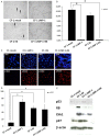
Apoptosis, DNA damage, and induction of DDR due to expression of EBVLMP-1, HPV16 E6, and LMP-1+E6. (A, B) MEFs expressing LMP-1, E6, or LMP-1+E6, or mock were cultured under serum-free conditions, and TUNEL was used to detect apoptotic cells. At least 200 cells were counted in each experiment. The arrows in (A) indicate apoptotic cells. Data in (B) are mean ± SDs from three independent experiments and are presented as TUNEL-positive cells/total cells. *P < 0.05. (C, D) γH2AX immunofluorescence in MEFs expressing LMP-1, E6, or LMP-1+E6, or mock. γH2AX positivity is an indicator of DNA damage. At least 200 cells were counted in each experiment. Data in (D) are mean ± SDs from three independent experiments and are presented as γH2AX-positive cells/total cells. *P < 0.05. (E) Analysis by Western blotting of p53, RB, Chk1, and p27 in MEFs expressing LMP-1, E6, or LMP-1+E6, or mock. β-actin was used as a loading control.
Figure 3
Figure 3
Anchorage-independent growth of CF-1 LMP-1+E6 cells. (A, B) MEFs expressing LMP-1, E6, LMP-1+E6, or mock were suspended in 0.7% agarose in 6-well plates (1 × 10⁴ cells/well), and colony formation was observed for 4 weeks. Arrows in (A) indicate colony formation. Magnification, 100 ×. The number of colonies was counted 4 weeks after plating. Data in (B) are the mean number of colonies ± SDs from three independent experiments.
Figure 4
Figure 4
Effects of LMP-1 or E6 gene knockdown on cell proliferation and anchorage-independent growth of CF-1 LMP-1+E6 cells. A: CF-1 LMP-1+E6 and CF-1 mock cells were transfected with siRNA for LMP-1, E6, or control siRNAs. The cells were analyzed by RT-PCR for expression of LMP-1, E6 and GAPDH mRNAs. B: CCK8 analysis of proliferation of CF-1 LMP-1+E6- and CF-1 mock cells transfected with LMP-1, E6, or control siRNAs. Data are means ± SDs from three independent experiments. C: Colony formation in soft agar by CF-1 LMP-1+E6 cells transfected with LMP-1, E6, or control siRNAs. Forty-eight hours after siRNA transfection, cells were suspended in 0.7% agarose and observed for 4 weeks for colony formation. The arrow indicates a colony. Magnification, 100 ×.
Figure 5
Figure 5
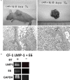
Tumor formation in nude mice after injection of CF-1 LMP-1+E6 cells. A: Large, lobulated tumors from the injection site on the back of the mice 61 days after injection. Magnification, 100 × (lower left panel) and 400 × (lower right panel). B: RT-PCR analysis of tumor mRNA, showing expression of LMP-1 and E6. Expression of GAPDH was used as a control. RT+ indicates that DNase-treated RNA was reacted with reverse transcriptase for cDNA synthesis, and RT- indicates DNase-treated RNA that was not reacted with reverse transcriptase to check genomic DNA contamination.
Figure 6
Figure 6
NF-ĸB activity in MEFs expressing LMP-1, and LMP-1+E6, and its suppression by knockdown of LMP-1 or E6. A: NF-ĸB activity relative to mock-transduced MEFs determined by luciferase assays in MEFs expressing EBV and/or HPV genes. CF-1 TNFα are MEFs treated with TNFα as a positive control. Data are means ± SDs from three independent experiments. *P < 0.05. B: Luciferase assays of NF-ĸB activities in CF-1 LMP-1, CF-1 LMP-1+E6 and CF-1 mock cells transfected with control or LMP-1- and E6-specific siRNAs. CF-1 TNFα represents MEFs treated with TNFα as a positive control. Data are means ± SDs from three independent experiments. *P < 0.05. **P < 0.01.

Similar articles

Cited by

References

    1. Raab-Traub N. Novel mechanisms of EBV-induced oncogenesis. Curr Opin Virol. 2012;2:453–458. - PMC - PubMed
    1. Suzuki R. NK/T-cell lymphomas: pathobiology, prognosis and treatment paradigm. Curr Oncol Rep. 2012;14:395–402. - PubMed
    1. Hammerschmidt W, Sugden B. Genetic analysis of immortalizing functions of Epstein-Barr virus in human B lymphocytes. Nature. 1989;340:393–397. - PubMed
    1. Kaye KM, Izumi KM, Kieff E. Epstein-Barr virus latent membrane protein 1 is essential for B-lymphocyte growth transformation. Proc Natl Acad Sci U S A. 1993;90:9150–9154. - PMC - PubMed
    1. Tomkinson B, Robertson E, Kieff E. Epstein-Barr virus nuclear proteins EBNA-3A and EBNA-3C are essential for B-lymphocyte growth transformation. J Virol. 1993;67:2014–2025. - PMC - PubMed

MeSH terms

Substances

LinkOut - more resources