

DCC is expressed in a CD166-positive subpopulation of chondrocytes in human osteoarthritic cartilage and modulates CRE activity
- PMID: 24966904
- PMCID: PMC4069887
DCC is expressed in a CD166-positive subpopulation of chondrocytes in human osteoarthritic cartilage and modulates CRE activity
Abstract
Objective: In a recent study we determined a strong differential expression of DCC in OA compared to normal chondrocytes and a strong impact of the DCC receptor on cellular mobility triggered by its ligand Netrin-1. Migration of chondrocytes or their progenitor cells may play a role in remodeling of cartilage and pathological conditions. The purpose of this study is to identify subsets of chondrocytes expressing DCC and to understand signaling pathways used by DCC in chondrocytes.
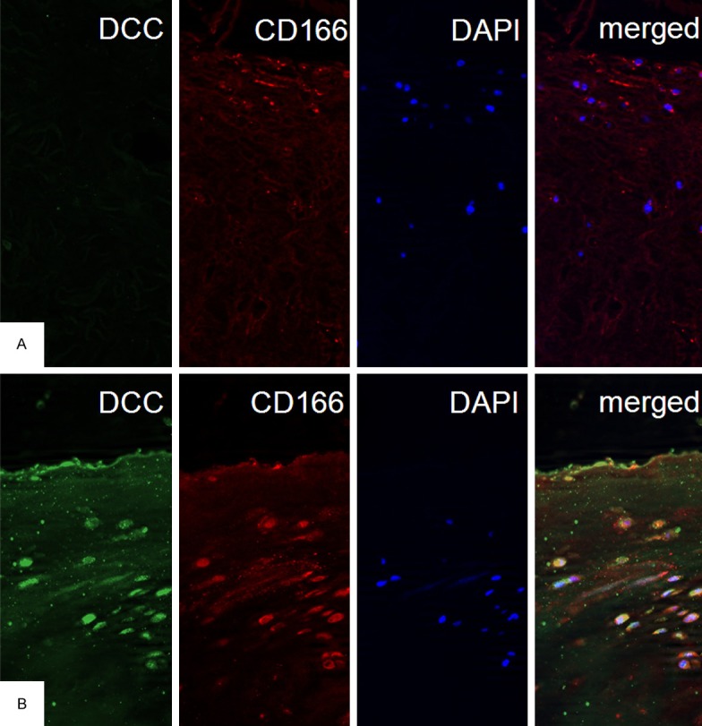
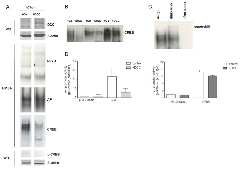
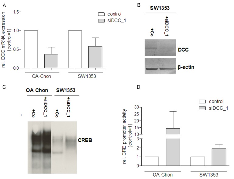
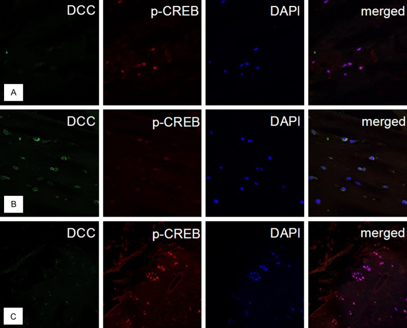
Methods: Immunofluorescent histology of human cartilage was used to determine the expression pattern of CD166, DCC and p-CREB. Cell culture of chondrocytes and SW1353, transient transfection, siRNA transfection, EMSA, luciferase assay, quantitative RT-PCR, ELISA, and Western Blotting were used to study signaling down-stream of DCC.
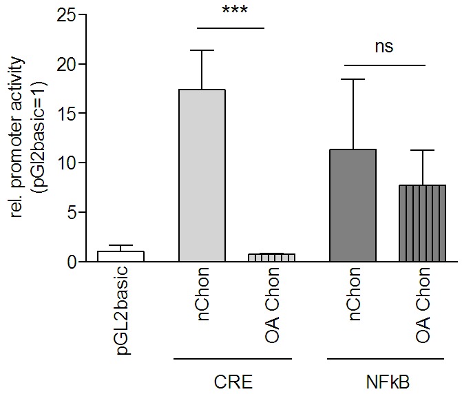
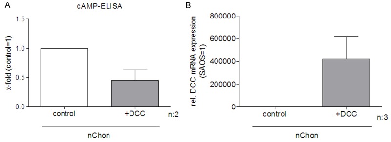
Results: DCC expressing chondrocytes are mainly located in the surface layers of OA cartilage. These also express CD166 indicating that DCC expressing chondrocytes are progenitor cells. Interestingly, expression of DCC reduces cAMP levels, CREB DNA-binding activity and CRE activity in chondrocytes, whereas down-regulation of DCC results in induction of CRE signaling.
Conclusion: In summary, DCC is up-regulated in CD166-positive chondrogenic progenitor cells in OA and induces down-regulation of CREB. These findings indicate that migration of CD166 positive progenitor cells to sites of cartilage damage may be directed by regulation of DCC signaling.
Keywords: CRE activity; CREB; DCC; Repellent factors; migration; osteoarthritis.
Figures
Similar articles
-
Role of deleted in colon carcinoma in osteoarthritis and in chondrocyte migration.Rheumatology (Oxford). 2009 Nov;48(11):1435-41. doi: 10.1093/rheumatology/kep245. Epub 2009 Sep 10. Rheumatology (Oxford). 2009. PMID: 19745029
-
Identification of mesenchymal progenitor cells in normal and osteoarthritic human articular cartilage.Arthritis Rheum. 2004 May;50(5):1522-32. doi: 10.1002/art.20269. Arthritis Rheum. 2004. PMID: 15146422
-
Relative percentage and zonal distribution of mesenchymal progenitor cells in human osteoarthritic and normal cartilage.Arthritis Res Ther. 2011 Apr 15;13(2):R64. doi: 10.1186/ar3320. Arthritis Res Ther. 2011. PMID: 21496249 Free PMC article.
-
Expression of functional mu-opioid receptors in human osteoarthritic cartilage and chondrocytes.Biochem Biophys Res Commun. 2003 Nov 7;311(1):202-7. doi: 10.1016/j.bbrc.2003.09.191. Biochem Biophys Res Commun. 2003. PMID: 14575714
-
[Experimental study on CD105+/CD166+ cells and its chondrogenic potential in early osteoarthritis cartilage].Zhongguo Xiu Fu Chong Jian Wai Ke Za Zhi. 2013 Jul;27(7):793-9. Zhongguo Xiu Fu Chong Jian Wai Ke Za Zhi. 2013. PMID: 24063165 Chinese.
Cited by
-
Research advances in cartilage stem cells markers and induced differentiation.Hua Xi Kou Qiang Yi Xue Za Zhi. 2021 Feb 1;39(1):108-114. doi: 10.7518/hxkq.2021.01.017. Hua Xi Kou Qiang Yi Xue Za Zhi. 2021. PMID: 33723946 Free PMC article. Review. Chinese, English.
-
ALCAM: A Novel Surface Marker on EpCAMlow Circulating Tumor Cells.Biomedicines. 2022 Aug 16;10(8):1983. doi: 10.3390/biomedicines10081983. Biomedicines. 2022. PMID: 36009530 Free PMC article.
-
Robo3A and Robo3B expression is regulated via alternative promoters and mRNA stability.Cancer Cell Int. 2016 Sep 20;16:71. doi: 10.1186/s12935-016-0347-9. eCollection 2016. Cancer Cell Int. 2016. PMID: 27660555 Free PMC article.
-
Cell Interplay in Osteoarthritis.Front Cell Dev Biol. 2021 Aug 3;9:720477. doi: 10.3389/fcell.2021.720477. eCollection 2021. Front Cell Dev Biol. 2021. PMID: 34414194 Free PMC article. Review.
-
Pathomechanisms of Posttraumatic Osteoarthritis: Chondrocyte Behavior and Fate in a Precarious Environment.Int J Mol Sci. 2020 Feb 25;21(5):1560. doi: 10.3390/ijms21051560. Int J Mol Sci. 2020. PMID: 32106481 Free PMC article. Review.
References
-
- Kim KW, Lim TH, Kim JG, Jeong ST, Masuda K, An HS. The origin of chondrocytes in the nucleus pulposus and histologic findings associated with the transition of a notochordal nucleus pulposus to a fibrocartilaginous nucleus pulposus in intact rabbit intervertebral discs. Spine (Phila Pa 1976) 2003;10:982–990. - PubMed
-
- Kim KW, Ha KY, Park JB, Woo YK, Chung HN, An HS. Expressions of membrane-type I matrix metalloproteinase, Ki-67 protein, and type II collagen by chondrocytes migrating from cartilage endplate into nucleus pulposus in rat intervertebral discs: a cartilage endplate-fracture model using an intervertebral disc organ culture. Spine (Phila Pa 1976) 2005;12:1373–1378. - PubMed
-
- Kambic HE, Futani H, McDevitt CA. Cell, matrix changes and alpha-smooth muscle actin expression in repair of the canine meniscus. Wound Repair Regen. 2000;6:554–561. - PubMed
Publication types
MeSH terms
Substances
LinkOut - more resources
Full Text Sources
Medical
Research Materials
Miscellaneous