. 2014 Apr 15;7(5):1967-76.
eCollection 2014.

Testosterone decreases the expression of endometrial pinopode and L-selectin ligand (MECA-79) in adult female rats during uterine receptivity period

Affiliations

Testosterone decreases the expression of endometrial pinopode and L-selectin ligand (MECA-79) in adult female rats during uterine receptivity period

Helmy Mohd Mokhtar et al. Int J Clin Exp Pathol. .

Abstract

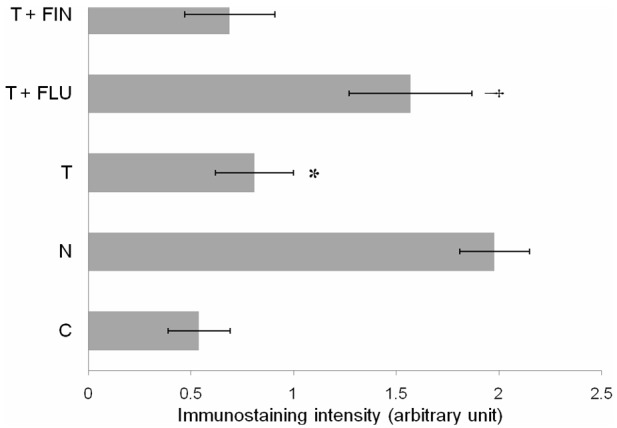
Pinopode, a progesterone-dependent endometrial projection which appears during uterine receptivity period, participates in blastocyst implantation. Blastocyst loosely attaches to pinopode via L-selectin ligand (MECA-79). We hypothesized that pinopode and MECA-79 expressions were affected by testosterone. Therefore, the effect of testosterone on pinopode and MECA-79 expressions during uterine receptivity period were investigated.

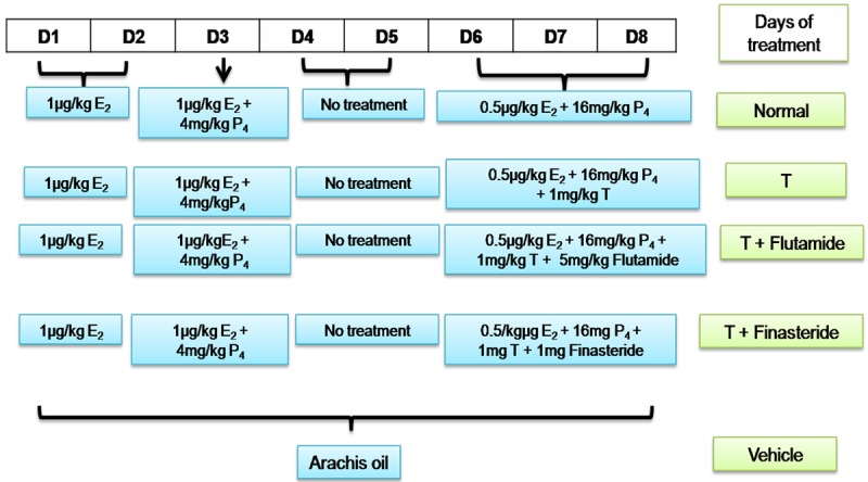
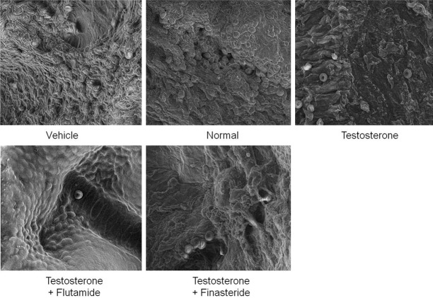
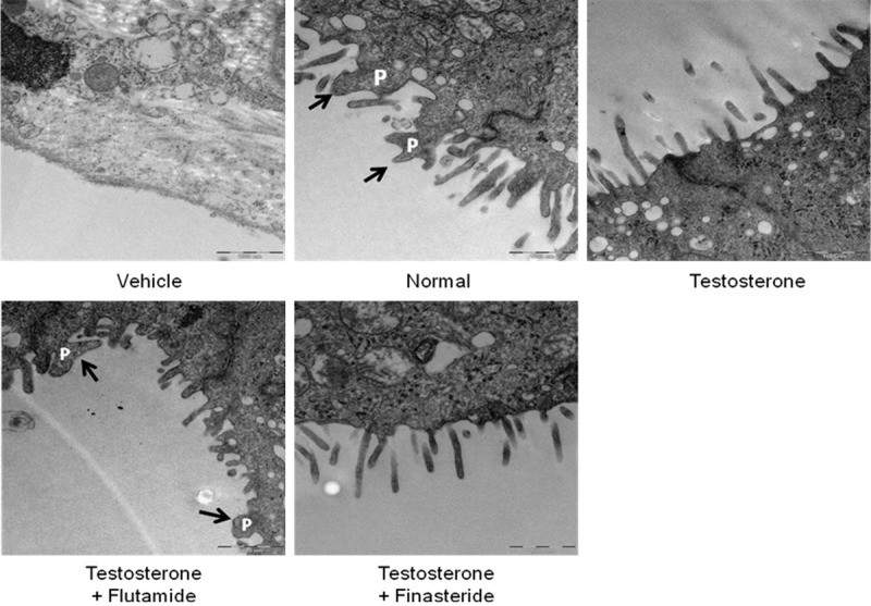
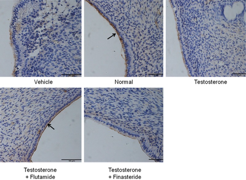
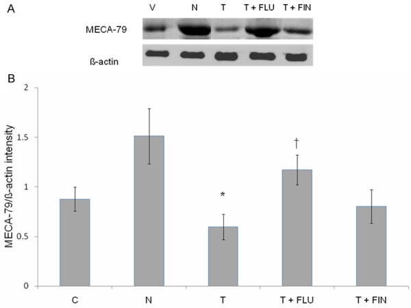
Methods: Ovariectomized adult female rats received 8 days sex-steroid replacement intended to mimic hormonal changes in early pregnancy with day 6 to 8 represents uterine receptivity period. Testosterone (1 mg/kg/day) was injected together with flutamide or finasteride during the period of uterine receptivity. At the end of treatment, rats were sacrificed and uteri were removed. The existence of pinopodes in the endometrium was visualized by electron microscopy and uterine expression and distribution of MECA-79 protein were examined by Western blotting and immunohistochemistry (IHC) respectively.

Results: Abundant pinopodes and MECA-79 expressions were observed in rats received normal steroid replacement regime. Administration of testosterone during uterine receptivity period reduced pinopodes and MECA-79 expressions, which were antagonized by flutamide and not finasteride.

Conclusions: The decrease in uterine pinopodes and MECA-79 expressions during uterine receptivity period by testosterone may cause failure of blastocyst to implant in conditions associated with high level of this hormone.

Keywords: MECA-79; Pinopode; testosterone; uterine receptivity.


Figures

Figure 1
Figure 1
Protocol of sex-steroid replacement regime. Schematic representation of time-course of steroid treatment protocol administered to ovariectomized rats to mimic the fluctuating steroid hormones level in early pregnancy. The animals were kept at 12 hours light and 12 hours dark schedule. D: day, T: testosterone.
Figure 2
Figure 2
Field emission scanning electron microscopy (FESEM) of the endometrium in different experimental groups. Abundant bleb-like projections were seen in rats received normal steroid-replacement regime. Fewer pinopodes were seen following administration of testosterone during uterine receptivity period which were reverted by flutamide but not finasteride. scale bar: 40 μM.
Figure 3
Figure 3
Transmission electron microscopy (TEM) of the endometrium in different experimental groups. Pinopode projections were seen in rats received normal steroid-replacement regime. Apical membrane with nearly absence pinopodes were seen following testosterone administration during uterine receptivity period which were reverted by flutamide but not finasteride. P = pinopode.
Figure 4
Figure 4
MECA-79 protein distribution at the apical membrane of the endometrial luminal epithelia in different experimental group. MECA-79 was seen to be distributed at the apical membrane of the luminal epithelia predominantly in rats received normal steroid replacement and concomitant testosterone and flutamide administration during uterine receptivity period. L: lumen, G: gland, scale bar: 50 μm. Arrow pointing towards the apical membrane with highest immunostaining intensity.
Figure 5
Figure 5
Quantitative analysis of MECA-79 immunoperoxidase staining intensity at the apical membrane of the luminal epithelia in different experimental groups. The highest intensity was observed in rats received normal steroid replacement regime and following co-administration of flutamide with testosterone during uterine receptivity period. Mean was obtained from intensity of 4 different sections per treatment group. *P < 0.05 as compared to normal steroid replacement, †P < 0.05 as compared with testosterone only treatment. C: vehicle control, N: normal steroid replacement regime, T: testosterone only injection with P4, T + FLU: testosterone and flutamide administration with P4 and T + FIN: testosterone and finasteride administration with P4.
Figure 6
Figure 6
Immunoblotting analysis of MECA-79 protein in different experimental group. (A) Shows immunoblot image of protein band in different experimental group while (B) Shows analyses of the intensity of protein bands in different experimental groups. Our findings indicate that the highest MECA-79 protein intensity was observed in the group received normal sex-steroid replacement while the lowest intensity was observed following administration of testosterone during the uterine receptivity period. Mean was obtained from intensity of 4 different membranes per treatment group. *P < 0.05 as compared to normal steroid replacement, †P < 0.05 as compared with testosterone only treatment. C: vehicle control, N: normal steroid replacement regime, T: testosterone only injection with P4, T + FLU: testosterone and flutamide administration with P4 and T + FIN: testosterone and finasteride administration with P4.

Similar articles

Cited by

References

    1. Martel D, Monier MN, Roche D, Psychoyos A. Hormonal dependence of pinopode formation at the uterine luminal surface. Hum Reprod. 1991;6:597–603. - PubMed
    1. Singh MM, Chauhan SC, Trivedi RN, Maitra SC, Kamboj VP. Correlation of pinopod development on uterine luminal epithelial surface with hormonal events and endometrial sensitivity in rat. Eur J Endocrinol. 1996;135:107–117. - PubMed
    1. Nikas G, Drakakis P, Loutradis D, Mara-Skoufari C, Koumantakis E, Michalas S, Psychoyos A. Implantation: Uterine pinopodes as markers of the ‘nidation window’ in cycling women receiving exogenous oestradiol and progesterone. Hum Reprod. 1995;10:1208–1213. - PubMed
    1. Ding T, Song H, Wang X, Khatua A, Paria BC. Leukemia inhibitory factor ligand-receptor signaling is important for uterine receptivity and implantation in golden hamsters (Mesocricetus auratus) Reproduction. 2008;135:41–53. - PubMed
    1. Aghajanova L, Hamilton AE, Giudice LC. Uterine receptivity to human embryonic implantation: Histology, biomarkers, and transcriptomics. Sem Cell Dev Biol. 2008;19:204–211. - PMC - PubMed

Publication types

LinkOut - more resources