Review
. 2014 Jan 1;8(1):1-12.
doi: 10.3941/jrcr.v8i1.1551. eCollection 2014 Jan.

Adrenal and extra-adrenal myelolipomas - a comparative case report

Affiliations
Review

Adrenal and extra-adrenal myelolipomas - a comparative case report

Arsany Hakim et al. J Radiol Case Rep. .

Abstract

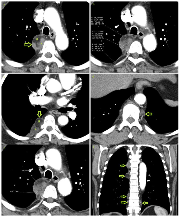
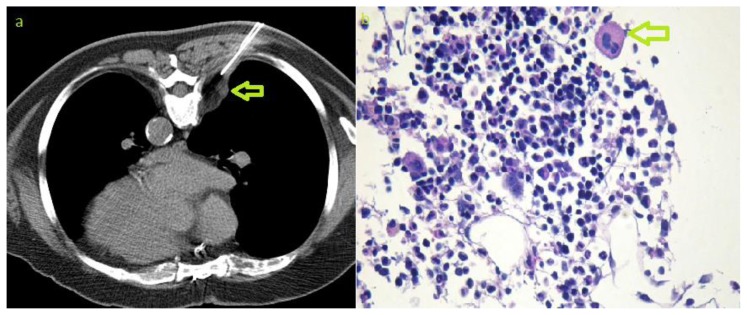
Myelolipoma is an uncommon benign tumour composed of mature fat tissue and haematopoietic elements and is most commonly found in the adrenal gland. We report a case, which was discovered incidentally on chest X-ray, of a rare occurrence of multifocal extra-adrenal myelolipoma in the thoracic paravertebral region. This was further investigated with multi-detector computed tomography and magnetic resonance imaging. The presumed diagnosis, of extra-adrenal myelolipoma, was histologically confirmed via tissue sample obtained by computed tomography guided biopsy. We compare the adrenal and extra-adrenal entities from the perspective of published literature and also review the cases, published in Pubmed, of extra-adrenal myelolipomas in order to summarize the different locations of this lesion.

Keywords: CT; Extra-adrenal; MRI; adrenal; fat; incidental; myelolipoma; paravertebral.


Figures

Figure 1
Figure 1
70-year-old male with bilateral multifocal paravertebral extra-adrenal myelolipoma. Chest X-ray showing incidentally detected right paravertebral mass (arrows). Plain chest radiography, posteranterior (a) and lateral (b), erect posture.
Figure 2
Figure 2
70-year-old male with bilateral multifocal extra-adrenal myelolipoma. Contrast enhanced CT of the chest in an arterial phase demonstrating a well-circumscribed right paravertebral mass at the level of the aortic arch measuring 45×26×56mm(a & e). The mass shows mixed density, partially hypodense with −70 hounsfield units (b) due to fat content (#) and partially hyperdense due to myeloid tissue (*). Multiple smaller lesions are shown bilaterally (arrows in c, d & f). No infiltration of the neighbouring tissues. (Technique: 16-MDCT scanner, Automatic tube current modulation ranging between 131 and 250 mA, 120KVp. 5mm slice thickness. Soft tissue window, level/width: 15/350. i.v. injection of 60ml of 300mg/ml iodine concentration non-ionic contrast. a)–e) axial plane, (f) coronal reformation).
Figure 3
Figure 3
70-year-old male with bilateral multifocal paravertebral extra-adrenal myelolipoma. MRI of the thoracic spine showing a right paravertebral mass with mixed signal intensity, partially hyperintense on T2 (a) and T1WI (b) due to the fat content of the mass (#) which shows signal drop-out on the fat suppression images (c & e) and on STIR sequence (f), and partially isointense on T2 (a) and hypointense on T1WI (b) due to the myeloid component (*) which shows hyperintensity on STIR (f), and moderate enhancement (c & e). Other similar lesions (arrows) are shown on the coronal plane (d). (Technique: 1.0 T scanner. a) axial T2WI: TR/TE: 3990/105, slice thickness 8, spacing 11.20, acquisition matrix: 256×205. b) axial T1WI: TR/TE: 509/11, slice thickness: 8, spacing 11.20, acquisition matric: 256×205. c) axial contrast enhanced T1WI with fat suppression: TR/TE: 742/12, slice thickness: 8, spacing: 11.20, acquisition matrix: 256×205. Administration of 15ml gadolinium based contrast agent. d) coronal non-enhanced T1WI: TR/TE: 863/13, slice thickness: 6, spacing 7.20, acquisition matrix: 187×512. e) coronal contrast enhanced T1WI with fat suppression: TR/TE: 1020/11, slice thickness: 6, spacing 7.20, acquisition matrix: 205×256. f) Sagittal Short tau inversion recovery (STIR): TR/TE: 3330/63, slice thickness: 3, spacing 3.60, acquisition matrix: 256×205)
Figure 4
Figure 4
70-year-old male with bilateral multifocal paravertebral extra-adrenal myelolipoma. CT-guided biopsy. a) axial non-enhanced CT obtained during biopsy procedure showing the needle that was advanced in a dorsal approach for biopsy of the paravertebral lesion (arrow). b) Photomicrography (Periodic acid Schiff stain, magnification 200x) showing bone marrow cells and megakaryocyte (arrow). Photomicrograph image courtesy of Prof. Alexander Tzankov, pathology department, University Hospital Basel, Switzerland)
Figure 5
Figure 5
(comparative case) 68-year-old female with left adrenal myelolipoma. Contrast enhanced CT of the abdomen in a venous phase showing a well-circumscribed mass in the region of the left adrenal gland measuring 86×99×95mm. The mass is composed of fat cells (#) with density of -109 HU(b) and myeloid tissue with relatively higher attenuation. The mass shows a pseudocapsule (arrows in a, c &d). No infiltration of the neighboring structures. (Technique: 16-MDCT scanner. Automatic tube current modulation ranging between 300 and 440 mA, 120 KVp, 5mm slice thickness. Soft tissue window, level/width: 40/400. i.v. injection of 100 ml Imeron 300. Coronal and sagittal reformatted CT with 5mm slice thickness. a) &b) axial planes, c) coronal reformation and d) sagittal reformation).

References

    1. Federle M, Anne V. Adrenal Myelolipoma. In: Federle M, editor. Diagnostic Imaging: Abdomen. 1st ed. Salt lake: Amirsys; 2004. pp. III 2 24–25.
    1. Gierke E. Uber Knochenmarksgewebe in der Nebenniere. Beitr Pathol Anat. 1905;7:311–25.
    1. Oberling C. Les formation myelo-lipomateuses. Bull Assoc Fr Etud Cancer. 1929;18:234–246.
    1. Rao P, Kenney PJ, Wagner BJ, Davidson AJ. Imaging and pathologic features of myelolipoma. Radiographics. 1997 Nov-Dec;17(6):1373–85. - PubMed
    1. Arzanian MT, Khaleghnejad-Tabari A, Shamsian BS, Jadali F, Gharib A, Esfahani H. Generalized myelolipoma. Arch Iran Med. 2006 Jul;9(3):274–6. - PubMed