Case Reports
. 2014 Jan 1;8(1):34-40.
doi: 10.3941/jrcr.v8i1.1424. eCollection 2014 Jan.

Management of iatrogenic urinothorax following ultrasound guided percutaneous nephrostomy

Affiliations
Case Reports

Management of iatrogenic urinothorax following ultrasound guided percutaneous nephrostomy

Deepak Batura et al. J Radiol Case Rep. .

Abstract

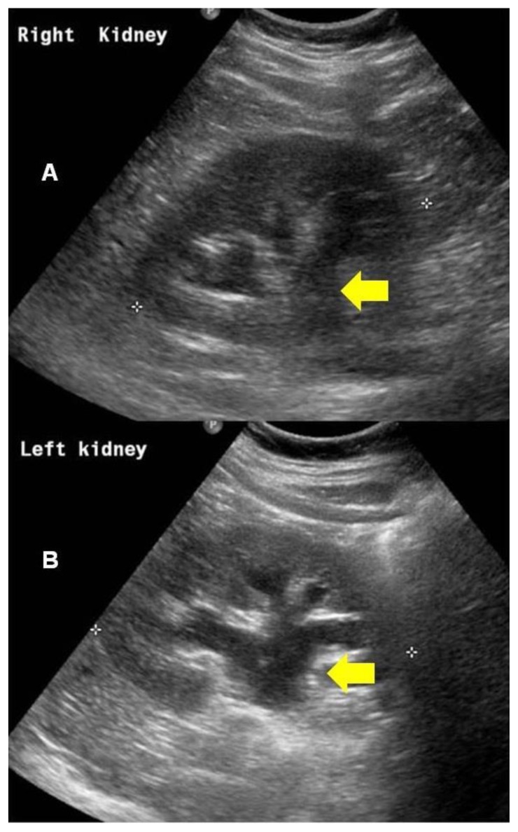
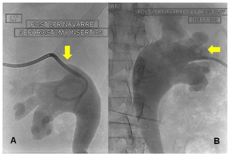
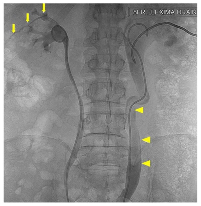
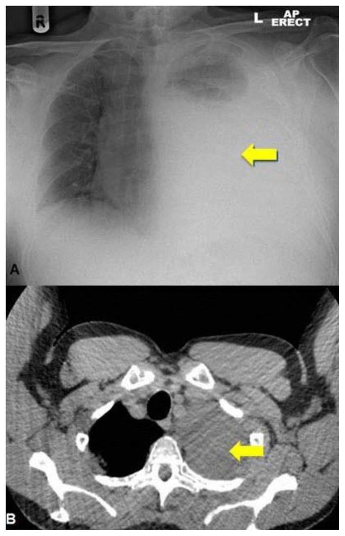
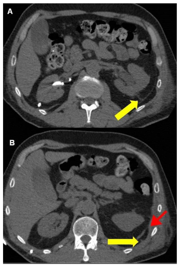
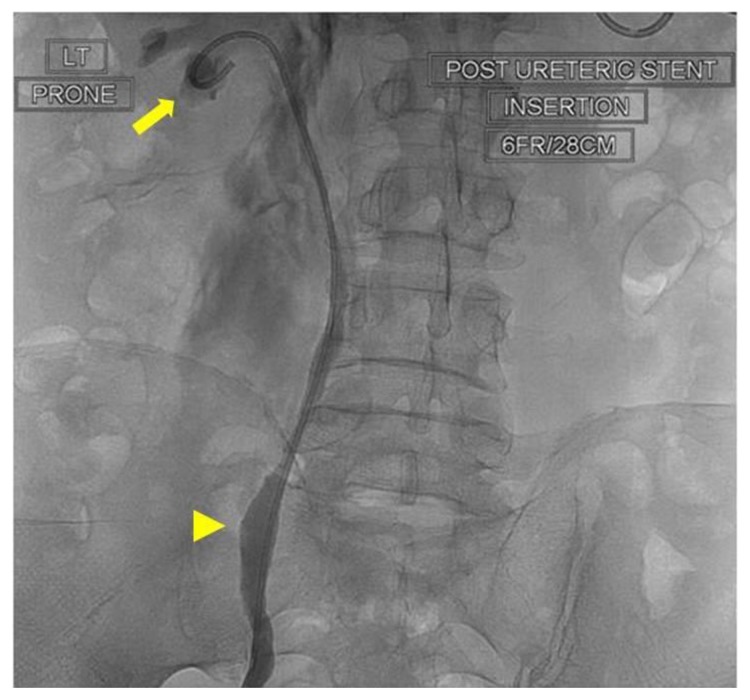
A 64 year-old male with metastatic prostate adenocarcinoma presented with bilateral hydronephrosis and renal impairment. Bilateral percutaneous nephrostomy drainage followed by ante-grade stenting was done. Shortly afterwards, the patient developed an extensive left-sided pleural effusion. His serum creatinine rose and he became anuric. Emergency pleural aspiration and later, pleural drainage were performed. Pleural aspirate was diagnostic of urinothorax and non contrast CT scan demonstrated a left reno-pleural fistula. The right stent was removed cystoscopically. The left stent could not be removed cystoscopically and was replaced in an ante grade manner through a fresh percutaneous renal approach. This led to cessation of pleural fluid accumulation. The patient was discharged with bilateral ureteric stents and normal renal function. A month later, he had normal renal function, no hydronephrosis and normal chest x-rays.

Keywords: Urinothorax; complication; percutaneous nephrostomy; prostate cancer; reno-pleural fistula; ureteral stent.


Figures

Figure 1
Figure 1
Right kidney (A) and left kidney (B). 64 year old male with an iatrogenic left urinothorax. Renal ultrasound scans showing bilateral moderate hydronephrosis secondary to bilateral ureteric obstruction by prostate cancer at bladder trigone. Arrows show dialted renal collecting systems. (Transabdominal scan using a 2–5MHz curvilinear probe).
Figure 2
Figure 2
64 year old male with an iatrogenic left urinothorax. Bilateral nephrostograms at initial nephrostomy placement with 8Fr Navarre nephrostomy tubes (patient prone). Note bilateral moderate hydroureteronephrosis and superior calyceal puncture on left (A). The puncture on the right is lower (B). Fluoroscopic images were obtained after the percutaneous injection of non-ionic water-soluble contrast material through the nephrostomy catheters.
Figure 3
Figure 3
64 year old male with an iatrogenic left urinothorax. Nephrostogram after initial antegrade stenting (patient prone). No covering nephrostomy has been placed after stenting on the left. Contrast can be seen extravasating from the left superior calyx (arrows) and a large clot lies within the lumen of the dilated right ureter (arrowheads). Spot fluoroscopic image was obtained after the anterograde injection of non-ionic water-soluble contrast material.
Figure 4
Figure 4
Chest X-ray (A) and thoracic CT (B). 64 year old male with an iatrogenic left urinothorax. Composite thoracic x-ray and CT images showing the large urinothorax (arrows) which has increased in the lower image (4B). Erect AP Chest x-ray. CT protocol: unenhanced volumetric CT scan of the chest in the axial plane using a Phillips Brilliance 64 slice CT scanner.
Figure 5
Figure 5
64 year old male with an iatrogenic left urinothorax. Non-contrast CT images showing the reno-pleural fistula. The track can be seen both in the retoperitoneum (arrow, A) in the thoracic cavity (yellow arrow, B). The red arrow in the lower image (B) points to the diaphragm where it attaches to the chest wall. (Protocol: Phillips Brilliance 64 slice CT scanner, 2 mm slice thickness, no intravenous, or oral contrast).
Figure 6
Figure 6
64 year old male with an iatrogenic left urinothorax. Nephrostogram showing the left stent (6Fr, 28cm) (arrowhead) replaced through a lower calyx (arrow). Spot fluoroscopic image was obtained after the percutaneous injection of non-ionic water-soluble contrast material through the fresh (lower pole calyceal) puncture.

References

    1. Garcia-Pachon E, Romero S. Urinothorax: a new approach. Curr Opin Pulm Med. 2006;12(4):259–63. - PubMed
    1. Lahiry SK, Alkhafaji AH, Brown AL. Urinothorax following blunt trauma to the kidney. J Trauma. 1978;18(8):608–10. - PubMed
    1. Amro O, Webb-Smith F, Sunderji S. Urinothorax: a rare complication of total abdominal hysterectomy. Obstet Gynecol. 2009;114(2 Pt 2):482–4. - PubMed
    1. Izzo L, Caputo M, De TG, Izzo P, Bolognese A, Basso L. Urinoma and urinothorax: report of a case. Am Surg. 2008;74(1):62–3. - PubMed
    1. Karkoulias K, Sampsonas F, Kaparianos A, Tsiamita M, Tsoukalas G, Spiropoulos K. Urinothorax: an unexpected cause of pleural effusion in a patient with non-Hodgkin lymphoma. Eur Rev Med Pharmacol Sci. 2007;11(6):373–4. - PubMed

Publication types

LinkOut - more resources