

Heterotopic ossification of the quadratus lumborum muscle
- PMID: 24967013
- PMCID: PMC4037244
- DOI: 10.3941/jrcr.v8i1.1348
Heterotopic ossification of the quadratus lumborum muscle
Abstract
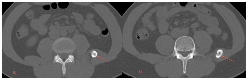
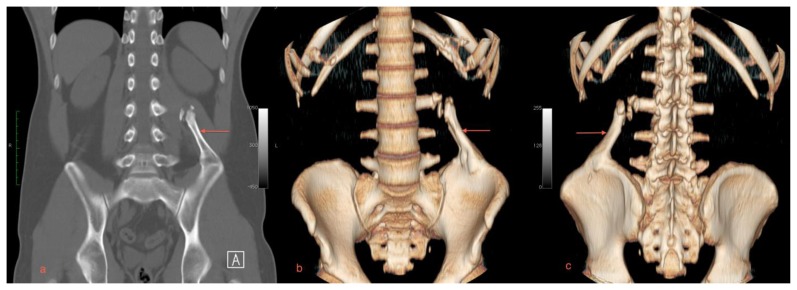
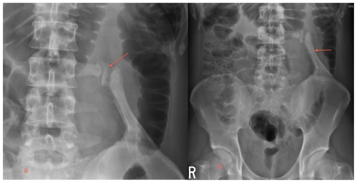
Heterotopic ossification is a benign process of mature laminar bone formation in the soft tissues. A synonymous term used to describe this pathology in muscle is myositis ossificans. The pathogenesis is unclear, but is likely multifactorial. The basic pathology is thought to be ectopic production of osseous tissue as part of a repair process in response to tissue injury. This report describes a case of heterotopic ossification of the quadratus lumborum muscle as an incidental finding. This case highlights that treatment is based on symptoms and conservative management might be appropriate for the asymptomatic patient.
Keywords: Quadratus lumborum muscle; computed tomography; idiopathic ossification; imaging.
Figures
Similar articles
-
Traumatic heterotopic ossification of the quadratus lumborum: a case report.Iowa Orthop J. 2008;28:88-90. Iowa Orthop J. 2008. PMID: 19223956 Free PMC article. No abstract available.
-
Myositis ossificans in psoas muscle after lumbar spine fracture.Spine (Phila Pa 1976). 2009 May 1;34(10):E367-70. doi: 10.1097/BRS.0b013e31819b30bf. Spine (Phila Pa 1976). 2009. PMID: 19404167
-
Heterotopic ossification: a review.Skeletal Radiol. 2005 Oct;34(10):609-19. doi: 10.1007/s00256-005-0958-z. Epub 2005 Aug 25. Skeletal Radiol. 2005. PMID: 16132978 Review.
-
Heterotopic Ossification in Mouse Models of Fibrodysplasia Ossificans Progressiva.Methods Mol Biol. 2019;1891:247-255. doi: 10.1007/978-1-4939-8904-1_18. Methods Mol Biol. 2019. PMID: 30414138 Free PMC article.
-
[Idiopathic pulmonary hemosiderosis with dendriform pulmonary ossification].Biomedica. 2016 Dec 1;36(4):504-508. doi: 10.7705/biomedica.v36i4.2968. Biomedica. 2016. PMID: 27992976 Review. Spanish.
Cited by
-
BMP-7 accelerates the differentiation of rabbit mesenchymal stem cells into cartilage through the Wnt/β-catenin pathway.Exp Ther Med. 2017 Dec;14(6):5424-5428. doi: 10.3892/etm.2017.5210. Epub 2017 Sep 27. Exp Ther Med. 2017. PMID: 29285071 Free PMC article.
References
-
- Vanden Bossche L, Vanderstraeten G. Heterotopic ossification: a review. J Rehabil Med. 2005 May;37(3):129–36. - PubMed
-
- Sawyer J, Myers M, Rosier R, Puzas JE. Heterotopic ossification: clinical and cellular aspects. Calcified Tissue International. 1991 Sep;49(3):208–15. - PubMed
-
- Mavrogenis AF, Soucacos PN, Papagelopoulos PJ. Heterotopic ossification revisited. Orthopedics. 2011;34(3):177. - PubMed
Publication types
MeSH terms
LinkOut - more resources
Full Text Sources
Other Literature Sources
Medical