Case Reports
. 2014 Feb 1;8(2):46-53.
doi: 10.3941/jrcr.v8i2.1489. eCollection 2014 Feb.

Testicular adrenal rest tumors in a patient with congenital adrenal hyperplasia

Affiliations
Case Reports

Testicular adrenal rest tumors in a patient with congenital adrenal hyperplasia

Jeffrey Dee Olpin et al. J Radiol Case Rep. .

Abstract

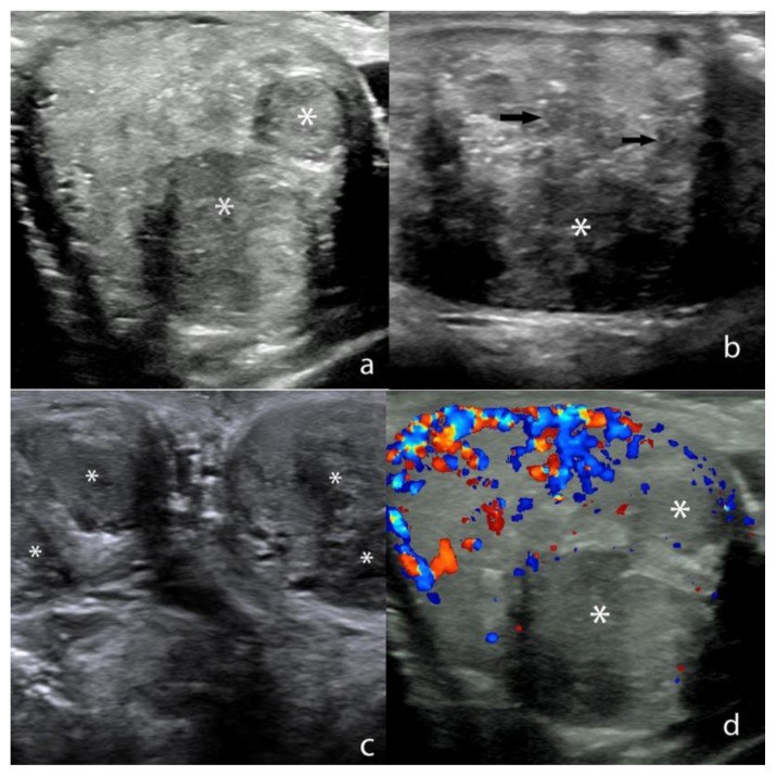
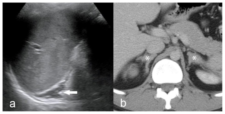
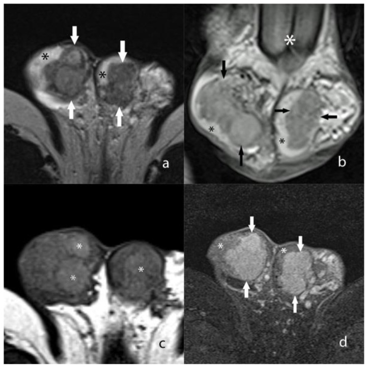
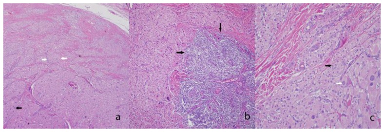
Congenital adrenal hyperplasia refers to a group of autosomal recessive disorders caused by a deficiency of an enzyme involved in the synthesis of glucocorticoids. The enzyme deficiency generally leads to a deficiency of cortisol and/or aldosterone production within the adrenal cortex. The lack of glucocorticoids generally leads to elevated levels of plasma corticotropin (ACTH), which often results in adrenal hyperplasia. Testicular adrenal rest tumors may develop in males with congenital adrenal hyperplasia due to overstimulation of aberrant adrenal cells within the testes. Recognition of this disease entity is essential when evaluating young males with testicular masses.

Keywords: Congenital adrenal hyperplasia; testicular adrenal rest tumors; testicular ultrasonography.


Figures

Figure 1
Figure 1
27 year old male with testicular adrenal rest tumors. a) Transverse Gray-scale ultrasound image through the right testis shows sharply marginated masses (*) that are hypoechoic relative to the normal background testicular parenchyma. Technique: 18 MHz high frequency linear-array transducer (Acuson). b) Sagittal Gray-scale ultrasound image through the left testis shows a dominant hypoechoic mass (*) with multiple other smaller, partially coalescent hypoechoic lesions scattered throughout the testis (arrows). Technique: 18 MHz high frequency linear-array transducer (Acuson). c) Transverse Gray-scale ultrasound image through both testes shows multiple bilateral, partially coalescent hypoechoic masses (*). Technique: 18 MHz high frequency linear-array transducer (Acuson). d) Transverse ultrasound image through the right testis with color Doppler interrogation shows hypoechoic masses (*) that show little to no vascular flow. Normal color Doppler flow is noted within the uninvolved testicular parenchyma. Technique: 18 MHz high frequency linear-array transducer (Acuson).
Figure 2
Figure 2
27 year old male with testicular adrenal rest tumors. a) Sagittal Gray-scale ultrasound image of the left upper quadrant shows diffuse, uniform enlargement of the left adrenal gland (arrow). Technique: 4.5 MHz vector transducer (Acuson). b) Contrast enhanced axial CT in the portal venous phase shows bilateral, symmetric enlargement of the adrenal glands (*). Technique: CT (Siemens Somatom) mA 380, kV 120, slice thickness 5 mm, 150 ml Isovue-300 IV contrast, portal venous phase.
Figure 3
Figure 3
27 year old male with testicular adrenal rest tumors. a) Axial T2 weighted MR image shows heterogeneous, coalescent masses (arrows) that occupy the majority of the testes. A small portion of the uninvolved hyperintense background testicular parenchyma is seen (*). Technique: 1.5 Tesla MRI (Siemens Avanto) TR/TE 5750/86, 5 mm slice thickness, non-contrast. b) Coronal T2 weighted MR image shows heterogeneous, coalescent masses (arrows) that are hypointense relative to the background testicular parenchyma (black asterisk). The base of the penis is seen on this image (white asterisk). Technique: 1.5 Tesla MRI (Siemens Avanto) TR/TE 5750/86, 5 mm slice thickness, non-contrast. c) Unenhanced axial T1 weighted MR image without fat saturation shows diffuse heterogeneity throughout both testes which contain discrete hyperintense foci (*). However, the outer tumor margins are not well visualized. Technique: 1.5 Tesla MRI (Siemens Avanto) TR/TE 514/17, 5 mm slice thickness, non-contrast. d) Axial T1 weighted MR image with fat saturation following intravenous gadolinium administration shows avidly enhancing masses (arrows) within both testes. A small portion of uninvolved testicular parenchyma is seen (*). Technique: 1.5 Tesla MRI (Siemens Avanto) TR/TE 514/15 with fat saturation, 3 mm slice thickness following 2 ml/kg Multihance, late arterial phase.
Figure 4
Figure 4
27 year old male with testicular adrenal rest tumors. a) Low power image showing a proliferation of nested neoplastic cells (white arrows) with abundant eosinophilic cytoplasm, endocrine atypia (black arrow), and with collagenous bands dividing the tumor nests (*). (Hematoxylin and eosin stained section, original magnification ×40). b) Medium power image showing a lymphoid aggregate associated with the tumor (black arrows). (Hematoxylin and eosin stained section, original magnification ×100.) c) High power view of the neoplastic cells with their abundant granular cytoplasm, and round nuclei, features resembling adrenal cortical tissue (black arrow). Again, note the extensive neuroendocrine atypia (white arrow). (Hematoxylin and eosin stained section, original magnification ×400.)

References

    1. Speiser PW, White PC. Congenital adrenal hyperplasia. The New England journal of medicine. 2003;349(8):776–788. - PubMed
    1. Therrell BL, Jr, Berenbaum SA, Manter-Kapanke V, Simmank J, Korman K, Prentice L, Gonzalez J, Gunn S. Results of screening 1.9 million Texas newborns for 21-hydroxylase-deficient congenital adrenal hyperplasia. Pediatrics. 1998;101(4 Pt 1):583–590. - PubMed
    1. Auchus RJ. Management of the adult with congenital adrenal hyperplasia. International journal of pediatric endocrinology. 2010;2010:614107. - PMC - PubMed
    1. Wilkins L. Macrogenitosomia precox associated with hyperplasia of the androgenic tissue of the adrenal and death from corticoadrenal insufficiency. Endocrinology. 1940;26:385–395.
    1. Avila NA, Shawker TS, Jones JV, Cutler GB, Jr, Merke DP. Testicular adrenal rest tissue in congenital adrenal hyperplasia: serial sonographic and clinical findings. AJR American journal of roentgenology. 1999;172(5):1235–1238. - PubMed

Publication types