

Vascular type Ehlers-Danlos Syndrome with fatal spontaneous rupture of a right common iliac artery dissection: case report and review of literature
- PMID: 24967021
- PMCID: PMC4037255
- DOI: 10.3941/jrcr.v8i2.1568
Vascular type Ehlers-Danlos Syndrome with fatal spontaneous rupture of a right common iliac artery dissection: case report and review of literature
Abstract
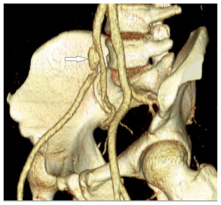
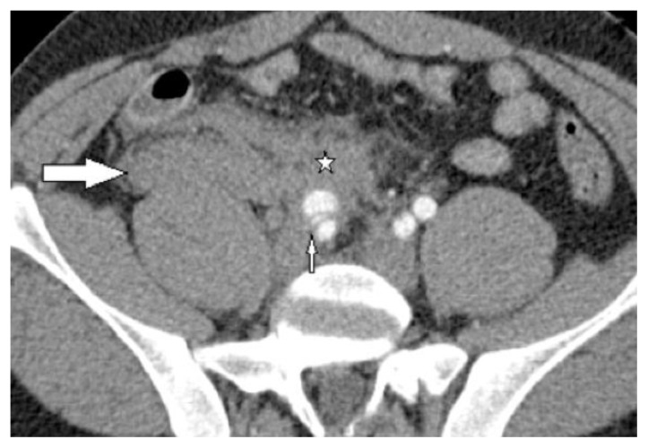
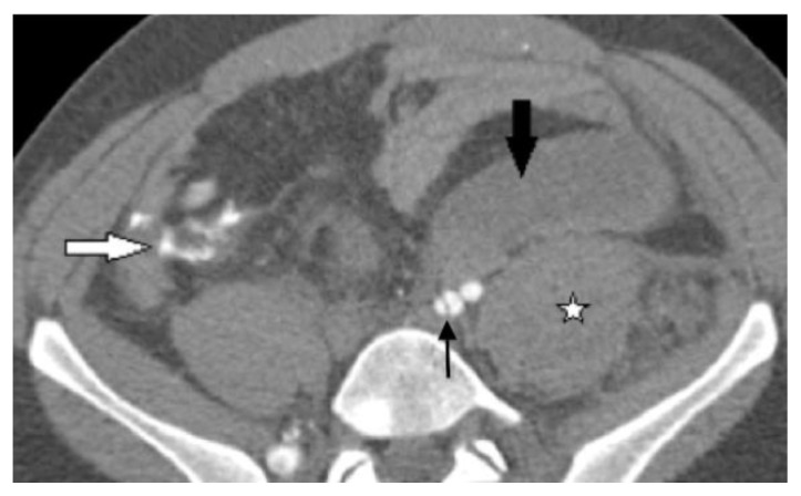
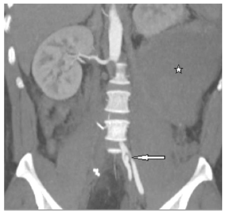
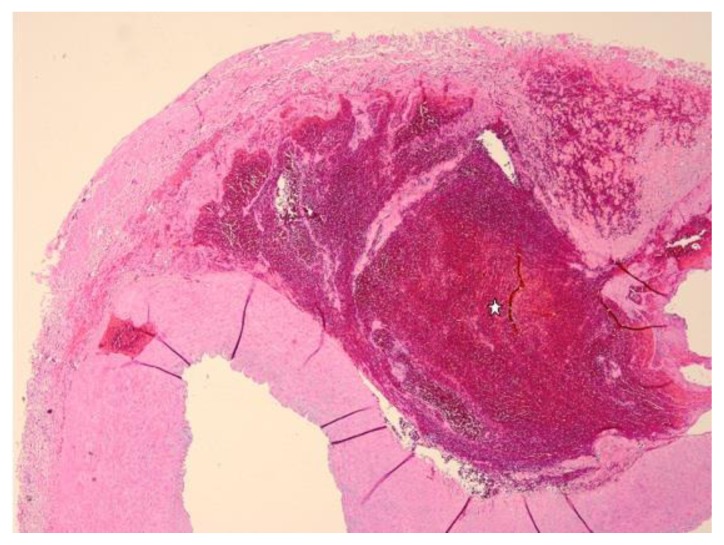
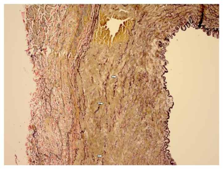
Vascular Ehlers-Danlos Syndrome (previously Ehlers-Danlos IV) is a rare autosomal dominant collagen vascular disorder caused by a 2q31 COL3A1 gene mutation encoding pro-alpha1 chain of type III collagen (in contrast to classic Ehlers-Danlos, caused by a COL5A1 mutation). The vascular type accounts for less than 4% of all Ehlers-Danlos cases and usually has a poor prognosis due to life threatening vascular ruptures and difficult, frequently unsuccessful surgical and vascular interventions. In 70% of cases, vascular rupture or dissection, gastrointestinal perforation, or organ rupture is a presenting sign. We present a case of genetically proven vascular Ehlers-Danlos with fatal recurrent retroperitoneal hemorrhages secondary to a ruptured right common iliac artery dissection in a 30-year-old male. This case highlights the need to suspect collagen vascular disorders when a young adult presents with unexplained retroperitoneal hemorrhage, even without family history of such diseases.
Keywords: Arterial dissection; Arterial rupture; Axial CT; Back pain; Collagen vascular disease; Common iliac artery dissection; Ectasia; Ehlers-Danlos Syndrome; Elastin; Hypovolemic shock; Iliac artery; Perforation; Retroperitoneal hematoma; Retroperitoneal hemorrhage; Vascular fragility; Vascular imaging; Vascular type Ehlers-Danlos Syndrome.
Figures
Similar articles
-
Peripartum Iliac Arterial Aneurysm and Rupture in a Patient with Vascular Ehlers-Danlos Syndrome Diagnosed by Next-Generation Sequencing.Int Heart J. 2018 Sep 26;59(5):1180-1185. doi: 10.1536/ihj.17-451. Epub 2018 Aug 29. Int Heart J. 2018. PMID: 30158381
-
Spontaneous mesenteric hemorrhage associated with Ehlers-Danlos syndrome.J Gastrointest Surg. 2006 Apr;10(4):583-5. doi: 10.1016/j.gassur.2005.07.018. J Gastrointest Surg. 2006. PMID: 16627225
-
Type IV Ehlers-Danlos syndrome with isolated arterial involvement.Ann Vasc Surg. 1990 Jan;4(1):15-9. doi: 10.1007/BF02042682. Ann Vasc Surg. 1990. PMID: 2297468
-
Presentation of Ehlers-Danlos syndrome: iliac artery pseudoaneurysm rupture.Ann Emerg Med. 1996 Aug;28(2):231-4. doi: 10.1016/s0196-0644(96)70066-5. Ann Emerg Med. 1996. PMID: 8759592 Review.
-
[Spontaneous rupture of common iliac artery: a case of Ehlers-Danlos syndrome and review of the literature].G Chir. 2006 Aug-Sep;27(8-9):324-7. G Chir. 2006. PMID: 17064493 Review. Italian.
Cited by
-
Spontaneous isolated iliac artery dissection in a young male: Case report and review of literature.Radiol Case Rep. 2024 Sep 10;19(12):5682-5687. doi: 10.1016/j.radcr.2024.08.064. eCollection 2024 Dec. Radiol Case Rep. 2024. PMID: 39308626 Free PMC article.
-
Spontaneous Isolated Dissection of Iliac Artery Treated with Endovascular Repair: A Case Report.Vasc Specialist Int. 2021 Dec 28;37:38. doi: 10.5758/vsi.210040. Vasc Specialist Int. 2021. PMID: 34961749 Free PMC article.
-
Anesthetic Management of a Patient With Ehlers-Danlos Syndrome.Anesth Prog. 2016 Winter;63(4):204-207. doi: 10.2344/16-00003.1. Anesth Prog. 2016. PMID: 27973938 Free PMC article.
-
Ehlers-Danlos Syndrome: Immunologic contrasts and connective tissue comparisons.J Transl Autoimmun. 2020 Dec 20;4:100077. doi: 10.1016/j.jtauto.2020.100077. eCollection 2021. J Transl Autoimmun. 2020. PMID: 33437956 Free PMC article. Review.
-
Spontaneous Dissection of the Renal Artery in Vascular Ehlers-Danlos Syndrome.Case Rep Crit Care. 2015;2015:804252. doi: 10.1155/2015/804252. Epub 2015 Jun 15. Case Rep Crit Care. 2015. PMID: 26175915 Free PMC article.
References
-
- Germain DP, Herrera-Guzman Y. Vascular Ehlers-Danlos syndrome. Ann Genet. 2004;47:1–9. - PubMed
-
- Germain DP. Clinical and genetic features of vascular Ehlers-Danlos syndrome. Ann Vasc Surg. 2002;16:391–397. - PubMed
-
- Superti-Furga A, Gugler E, Gitzelmann R, Steinman B. Ehlers-Danlos syndrome type IV: A multi-exon deletion in one of the two COL3A1 alleles affecting structures, stability, and processing of type III pro-collagen. J Biol Chem. 1988;1988;263:6226–6232. - PubMed
-
- Byers PH, Holbrook KA, McGillivray B, et al. Clinical and ultrastructural heterogeneity of type IV Ehlers-Danlos syndrome. Hum Genet. 1979;47:141–150. - PubMed
-
- Pope FM, Nicholls AC, Narcisi P, et al. Type III collagen mutations in Ehlers-Danlos syndrome type IV and other related disorders. Clin Exp Dermatol. 1988;13:285–302. - PubMed
Publication types
MeSH terms
LinkOut - more resources
Full Text Sources
Other Literature Sources
Medical
Miscellaneous