

Incidental intraosseous pneumatocyst with gas-density-fluid level in an adolescent: a case report and review of the literature
- PMID: 24967024
- PMCID: PMC4035364
- DOI: 10.3941/jrcr.v8i3.1540
Incidental intraosseous pneumatocyst with gas-density-fluid level in an adolescent: a case report and review of the literature
Abstract
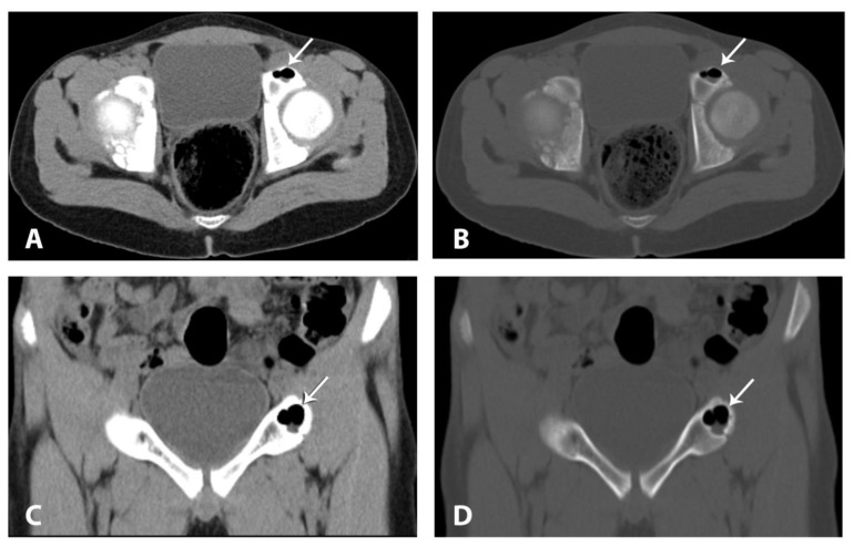
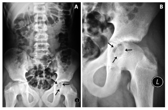
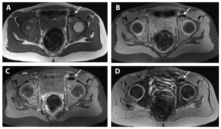
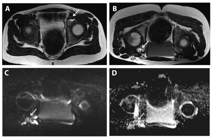
Intraosseous pneumatocyst is a gas containing lesion located within a bone. It is a relatively rare condition of unclear etiology and with an undetermined natural course. Gas-density-fluid level pneumatocyst is even rarer. Pneumatocyst is frequently seen in adults but rarely reported in pediatrics. The lesion is usually small and is seen in the vertebral bodies as well as around the sacroiliac joints. Rarely does it occur in other parts of the skeleton. We are reporting a case of large blood signal intensity containing intraosseous pneumatocyst in a 14 year old boy and reviewing other pediatric cases of pneumatocysts as well as those with gas-density-fluid level. The recognition of this incidental rare benign lesion is essential to avoid over investigation and an inappropriate aggressive intervention.
Keywords: CT; Gas-density-fluid level; Intraosseous pneumatocyst; MRI; Pediatrics.
Figures
Similar articles
-
Acetabular pneumatocyst containing air-fluid level.Eur Radiol. 1999;9(8):1647-9. doi: 10.1007/s003300050902. Eur Radiol. 1999. PMID: 10525883
-
Vertebral pneumatocyst. A case report.Spine (Phila Pa 1976). 1996 Feb 1;21(3):389-91. doi: 10.1097/00007632-199602010-00028. Spine (Phila Pa 1976). 1996. PMID: 8742219
-
Spontaneous progression of vertebral intraosseous pneumatocysts to fluid-filled cysts.Skeletal Radiol. 2001 Sep;30(9):523-6. doi: 10.1007/s002560100367. Skeletal Radiol. 2001. PMID: 11587521
-
Intraosseous pneumatocyst of the ilium: CT findings in two cases and literature review.Eur Radiol. 1997;7(9):1449-51. doi: 10.1007/s003300050315. Eur Radiol. 1997. PMID: 9369513 Review.
-
Vertebral body pneumatocyst in the cervical spine and review of the literature.Turk Neurosurg. 2008 Apr;18(2):197-9. Turk Neurosurg. 2008. PMID: 18597238 Review.
Cited by
-
Gas Bubbles in the Bone: A Case Report.J Clin Diagn Res. 2016 Jul;10(7):RD01-2. doi: 10.7860/JCDR/2016/19482.8200. Epub 2016 Jul 1. J Clin Diagn Res. 2016. PMID: 27630918 Free PMC article.
-
Axial Spondyloarthritis: Mimics and Pitfalls of Imaging Assessment.Front Med (Lausanne). 2021 Apr 22;8:658538. doi: 10.3389/fmed.2021.658538. eCollection 2021. Front Med (Lausanne). 2021. PMID: 33968964 Free PMC article. Review.
-
Prospective Evaluation of Magnetic Resonance Imaging Features of Magnesium-Based Alloy Screw Resorption in Pediatric Fractures.J Clin Med. 2023 Apr 21;12(8):3016. doi: 10.3390/jcm12083016. J Clin Med. 2023. PMID: 37109351 Free PMC article.
-
Intraosseous ganglion cyst mimicking chondrosarcoma on MRI: a case report.Eur J Med Res. 2022 Jan 13;27(1):8. doi: 10.1186/s40001-022-00631-0. Eur J Med Res. 2022. PMID: 35027087 Free PMC article.
-
Hip bone osteonecrosis with intraosseous pneumatosis after abdominal aortic aneurysm repair: a case of emphysematous osteomyelitis.BJR Case Rep. 2020 Oct 22;7(1):20200138. doi: 10.1259/bjrcr.20200138. eCollection 2021 Feb 1. BJR Case Rep. 2020. PMID: 33614123 Free PMC article.
References
-
- Fitzek S, Engelmann C, Fitzek C. Vertebral Pneumatization. Clin Neuroradiol. 2011;21:27–30. - PubMed
-
- Ramirez H, Blatt ES, Cable HF, McComb BL, Zornoza J, Hibri NS. Intraosseous Pneumatocyst of the ilium findings on radiographs and CT scans. Radiology. 1984;150:503–505. - PubMed
-
- Steingruber IE, Bach CM, Wimmer C, Nogler M, Buchberger W. Multisegmental pneumatocysts of lumbar spine mimic osteolytic lesions. Eur Radiol. 2001;11:845–848. - PubMed
-
- Narvμez JA, Narvμez J, Rodríguez-Mijarro1 M, Quintero JC. Acetabular pneumatocyst containing air-fluid level. Eur Radiol. 1999;9:1647–1649. - PubMed
Publication types
MeSH terms
Substances
LinkOut - more resources
Full Text Sources
Other Literature Sources