

Inflammatory pseudotumor of the liver: a rare case of recurrence following surgical resection
- PMID: 24967025
- PMCID: PMC4035365
- DOI: 10.3941/jrcr.v8i3.1459
Inflammatory pseudotumor of the liver: a rare case of recurrence following surgical resection
Abstract
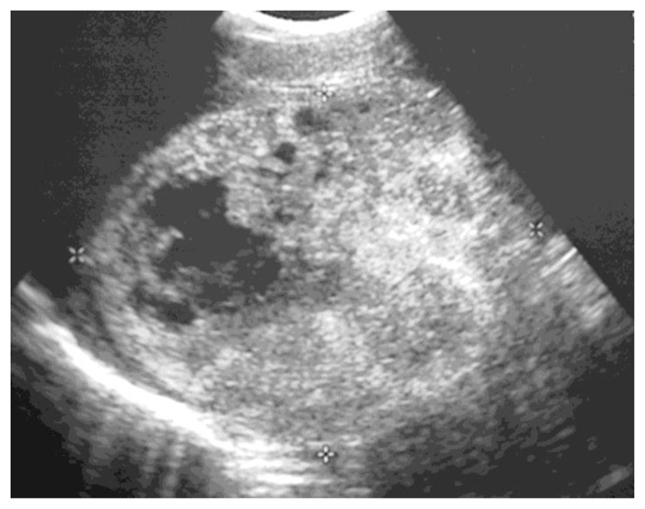
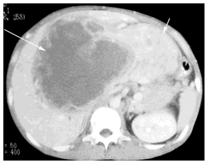
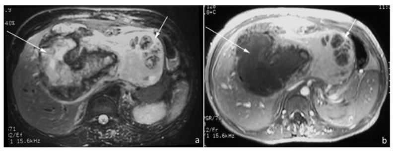
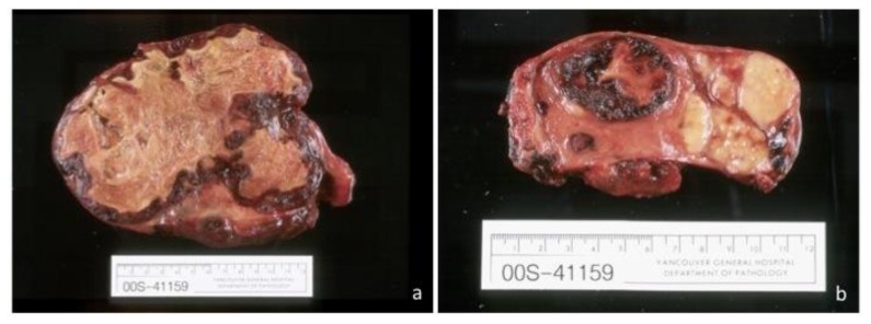
Inflammatory pseudotumor (IPT) of the liver is a rare, benign lesion that may be mistaken for malignancy. IPTs are difficult to diagnose due to non-specific clinical, laboratory and imaging features. We report the case of a 38-year old Asian male who presented with fatigue, weight loss and hepatomegaly. He was found to have a large hepatic IPT and underwent surgical resection; approximately two and a half years later, he developed acute cholangitis secondary to IPT recurrence. We present the imaging features of hepatic IPT using ultrasound, computed tomography (CT) and magnetic resonance imaging (MRI). We also review the literature on the diagnosis and management of this disease. The unique features of this case include the IPT's recurrence following surgical resection, large size and multiple modalities presented.
Keywords: Inflammatory pseudotumor; computed tomography; inflammatory myofibroblastic tumor; liver; magnetic resonance imaging; ultrasound.
Figures
Similar articles
-
Inflammatory pseudotumor of the liver treated by hepatic resection: a case report.Yonsei Med J. 2006 Feb 28;47(1):140-3. doi: 10.3349/ymj.2006.47.1.140. Yonsei Med J. 2006. PMID: 16502497 Free PMC article.
-
Hepatic inflammatory pseudotumor presenting in an 8-year-old boy: A case report and review of literature.World J Gastroenterol. 2015 Jul 28;21(28):8730-8. doi: 10.3748/wjg.v21.i28.8730. World J Gastroenterol. 2015. PMID: 26229415 Free PMC article. Review.
-
Hepatic inflammatory pseudotumor associated with xanthogranulomatous cholangitis mimicking cholangiocarcinoma.Intern Med. 2015;54(7):771-5. doi: 10.2169/internalmedicine.54.2623. Epub 2015 Apr 1. Intern Med. 2015. PMID: 25832940 Review.
-
Inflammatory pseudotumor of the liver and spleen diagnosed by percutaneous needle biopsy.World J Gastroenterol. 2012 Jan 7;18(1):90-5. doi: 10.3748/wjg.v18.i1.90. World J Gastroenterol. 2012. PMID: 22228976 Free PMC article.
-
[Inflammatory pseudotumors of the liver. Apropos of a pediatric case with radiological and ultrasonographic features and anatomopathological correlations].J Radiol. 1998 Jun;79(6):553-6. J Radiol. 1998. PMID: 9757283 French.
Cited by
-
Inflammatory Pseudotumor of the Liver or Intrahepatic Cholangiocarcinoma, That's the Question: A Review of the Literature.Cancers (Basel). 2024 Aug 23;16(17):2926. doi: 10.3390/cancers16172926. Cancers (Basel). 2024. PMID: 39272784 Free PMC article. Review.
-
Reactive lymphoid hyperplasia of the liver mimicking hepatocellular carcinoma: incidental finding of two cases.Int J Clin Exp Pathol. 2015 May 1;8(5):5863-9. eCollection 2015. Int J Clin Exp Pathol. 2015. PMID: 26191310 Free PMC article.
-
Multiple hepatic inflammatory pseudotumors with elevated alpha-fetoprotein and alpha-fetoprotein lectin 3 fraction with various PET accumulations: a case report.Surg Case Rep. 2021 Apr 28;7(1):107. doi: 10.1186/s40792-021-01188-6. Surg Case Rep. 2021. PMID: 33913027 Free PMC article.
-
Inflammatory Pseudotumor of the Liver Presented in a Patient with Cholelithiasis.Cureus. 2018 Aug 29;10(8):e3231. doi: 10.7759/cureus.3231. Cureus. 2018. PMID: 30410837 Free PMC article.
-
Clinicopathologic features and outcomes of hepatic inflammatory pseudotumour (IPT) and hepatic IPT-like lesions.Histopathology. 2025 Mar;86(4):525-535. doi: 10.1111/his.15357. Epub 2024 Oct 31. Histopathology. 2025. PMID: 39478419 Free PMC article.
References
-
- Das Narla L, Newman B, Spottswood S, Narla S, Kolli R. Inflammatory Pseudotumors. RadioGraphics. 2003;23:719–729. - PubMed
Publication types
MeSH terms
LinkOut - more resources
Full Text Sources
Other Literature Sources
Medical